Data processing method for low-frequency ultrasonic thoracic cavity imaging

A technology of data processing and thoracic cavity, applied in ultrasonic/sonic/infrasound image/data processing, ultrasonic/sonic/infrasonic diagnosis, ultrasonic/sonic/infrasonic Permian technology, etc., to reduce mean square error and improve signal-to-noise the effect of

Pending Publication Date: 2021-05-07
SHENYANG POLYTECHNIC UNIV
View PDF0 Cites 6 Cited by
  • Summary
  • Abstract
  • Description
  • Claims
  • Application Information

AI Technical Summary

Problems solved by technology

[0004] The purpose of the present invention is to provide a data processing method for low-frequency ultrasound thoracic imaging to solve some problems in the existing low-frequency ultrasound thoracic imaging technology, and to apply a combination of one ultrasonic probe to transmit and multiple ultrasonic probes to form a receiving array The transmit-receive mode can improve the signal-to-noise ratio of the data received by the array and reduce the mean square error of the data

Method used


Image

  • Data processing method for low-frequency ultrasonic thoracic cavity imaging
  • Data processing method for low-frequency ultrasonic thoracic cavity imaging
  • Data processing method for low-frequency ultrasonic thoracic cavity imaging

Examples


Embodiment Construction

[0027] The following will clearly and completely describe the technical solutions in the embodiments of the present invention with reference to the accompanying drawings in the embodiments of the present invention. Obviously, the described embodiments are only some, not all, embodiments of the present invention. Based on the embodiments of the present invention, all other embodiments obtained by persons of ordinary skill in the art without making creative efforts belong to the protection scope of the present invention.

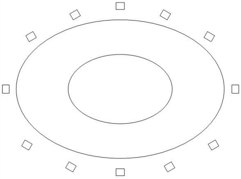
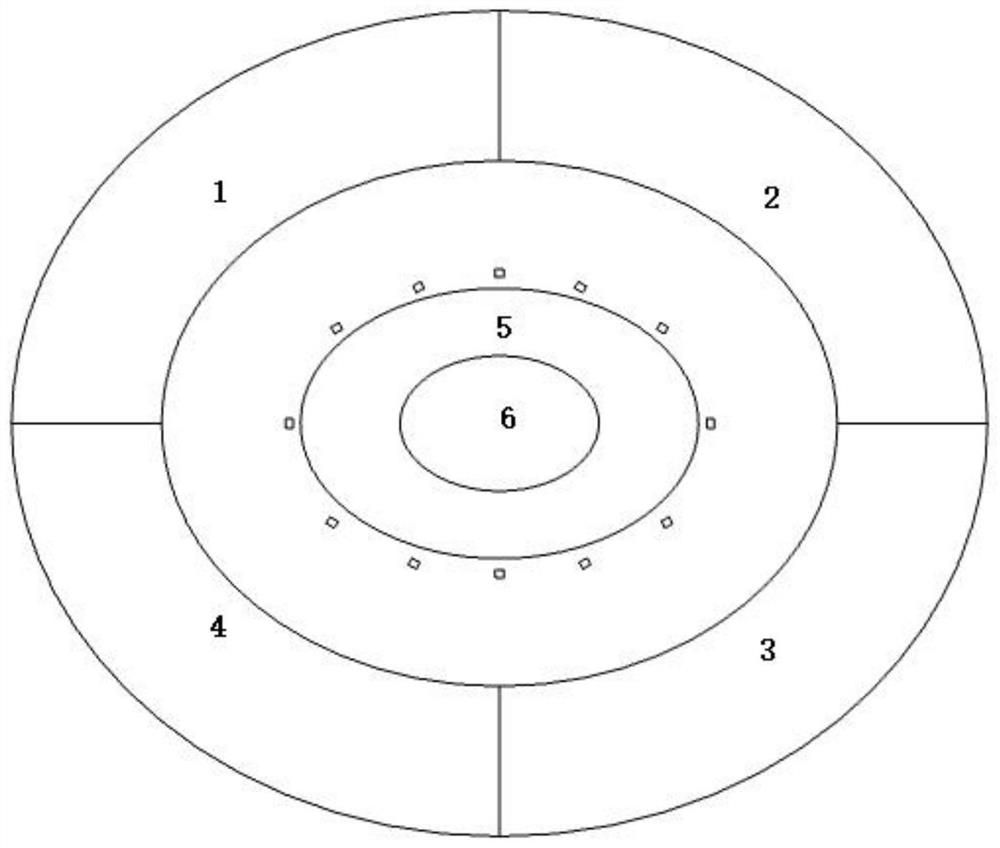
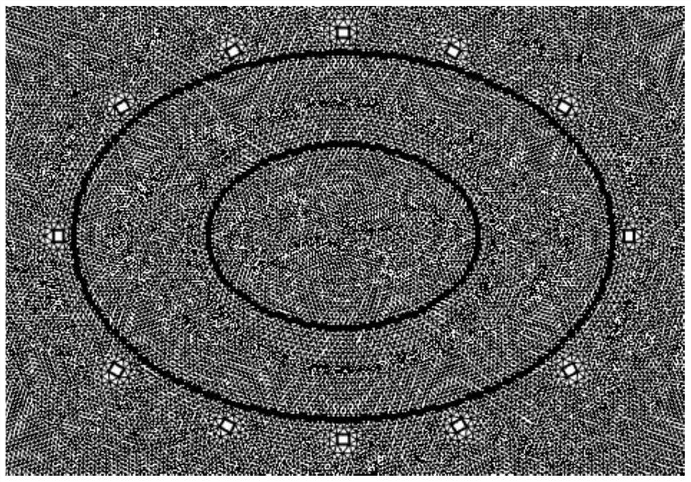
[0028] see Figure 1-11 , the present invention provides a technical solution: a data processing method for low-frequency ultrasound thoracic imaging, comprising

[0029] Modeling

[0030] Modeling is carried out through COMSOL, the spine and heart are ignored in the model, and the lungs are merged into an ellipse with a long axis of 0.15m and a short axis of 0.1m, and a ring-shaped skeletal muscle and skeleton are set outside the lung ellipse 12 ultrasonic ...


PUM


Abstract

The invention discloses a data processing method for low-frequency ultrasonic thoracic cavity imaging, which comprises the following steps: modeling through COMSOL, neglecting a spine and a heart in a model, combining two lungs into an ellipse with a long axis length of 0.15 m and a short axis length of 0.1 m, arranging a circle of annular skeletal muscle outside the ellipse of the lungs, uniformly placing 12 ultrasonic transducers around the exterior of the skeletal muscle, the material is set to be PZT-5H; one transducer is used as a sound source Sk which needs to be identified, seven transducers which directly face each other are used as receiving array elements, and signals received by the array elements are subjected to sound source identification processing by using a multi-signal classification (Multiple Signal Characterization,MUSIC) algorithm; a model is established through COMSOL, and a data processing technology applied to the field of low-frequency ultrasonic thoracic cavity imaging is creatively discovered according to a cyclic transmitting-receiving mode of one transmitting and seven receiving. According to the technology, the signal-to-noise ratio of array receiving data can be improved, and the mean square error of the data is reduced.

Description

technical field [0001] The invention relates to the technical field of low-frequency ultrasonic chest cavity imaging, and relates to a processing method for data received by an array. Background technique [0002] At present, traditional medical ultrasound imaging technology has become one of the main methods of in vivo imaging due to its non-invasiveness and non-radiation, but because the ultrasound used in traditional ultrasound imaging technology is in the range of 2-10MHz, ultrasound in this frequency band cannot penetrate Thoracic cavity, not available for thoracic imaging. Low-frequency ultrasound imaging technology is different from traditional ultrasound imaging. The ultrasound frequency emitted by it is between 20k-100k, which can penetrate the chest cavity and can be used for chest cavity imaging. [0003] Low-frequency ultrasound thoracic imaging technology uses transmission ultrasound data for imaging. The development of this technology is still in the prelimina...

Claims


Application Information

Patent Timeline
Patent Type & Authority Applications(China)
IPC IPC(8): A61B8/08A61B8/00
CPCA61B8/5207
Inventor 周英钢李运杰颜华罗浩梁凯张莺露
Owner SHENYANG POLYTECHNIC UNIV
Why Eureka
Try Eureka