Method for the preparation of biological, cytological, histological and autopsical samples and composition for mounting microscope slides

A compositional, cytological technique used in the preparation of test samples, microscopy, optics, etc.

Inactive Publication Date: 2021-05-07
戴尔帕斯有限公司
View PDF21 Cites 1 Cited by
  • Summary
  • Abstract
  • Description
  • Claims
  • Application Information

AI Technical Summary

Problems solved by technology

Such labels, required for identification, can be a source of failure for automatic coverslip clasps, as glue residue from the label often accumulates on the clasp, requiring xylene cleaning

Method used


Image

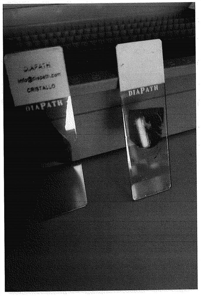
  • Method for the preparation of biological, cytological, histological and autopsical samples and composition for mounting microscope slides
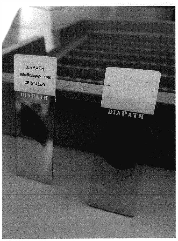
  • Method for the preparation of biological, cytological, histological and autopsical samples and composition for mounting microscope slides
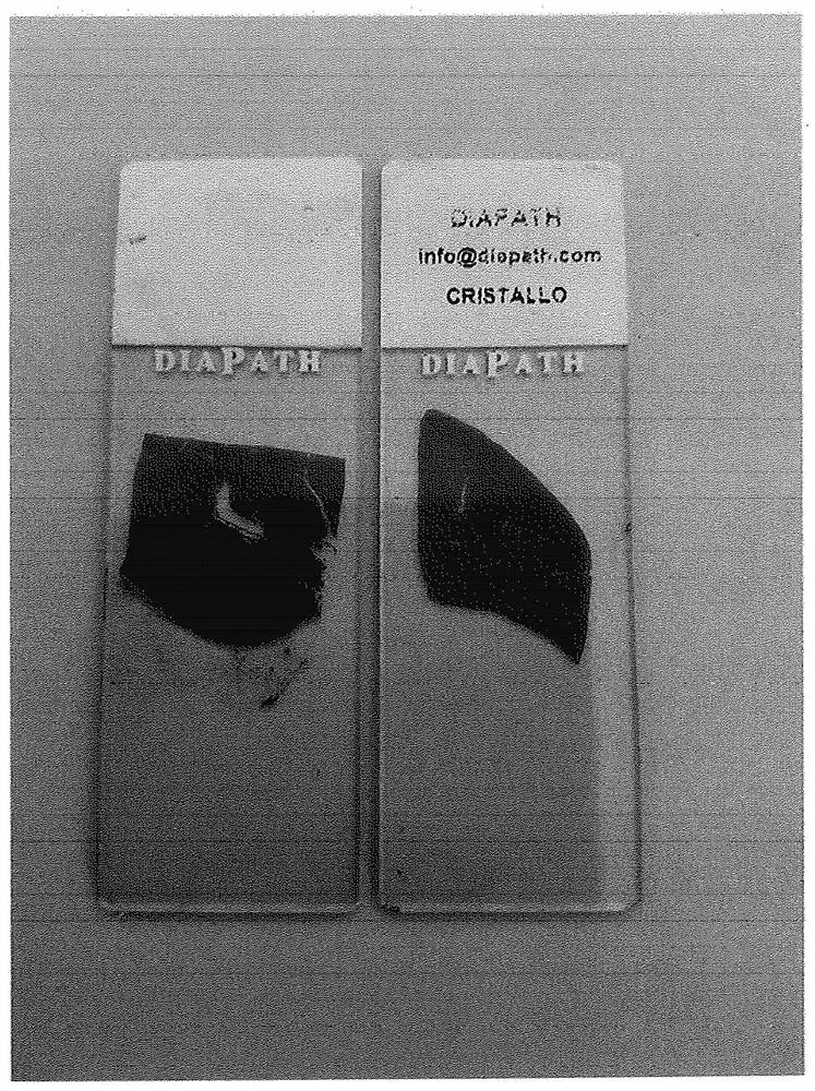
  • Method for the preparation of biological, cytological, histological and autopsical samples and composition for mounting microscope slides

Examples


Embodiment 1

[0128] Method for the preparation of one embodiment of the composition for use in the method of the invention

[0129] A suitable container (preferably opaque or amber) is charged with 750 ml of 1-methyl-2-methoxyethyl acetate. The container preferably has a lid so that no solvent is lost by evaporation.

[0130] 250 grams of cellulose acetate butyrate (CAB) was added to the solvent and the mixture was left under stirring for about 30 minutes until a homogeneous clear mixture was obtained. The process was carried out entirely at room temperature, with mechanical stirring stopped only when a homogeneous, clear mixture free of particulate matter was obtained.

Embodiment 2

[0132] Embedded method

Embodiment 21

[0133] Embodiment 2.1 (comparative example)

[0134] Conventional Manual Immobilization Process

[0135]At the end of the usual manual staining process that ends with alcohol washes, each of the 30 microscope slides undergoes the following steps: (I) place the microscope slide horizontally on the bench; (II) apply an appropriate amount of mounting medium Delivery onto microscope slide (about 2 drops); (III) placement of coverslip; and (IV) conditioning of coverslip. Total time required to immobilize each microscope slide: about 12 seconds; multiplied by each microscope slide: about 360 seconds (ie, about 6 minutes).

[0136] To the total time taken for routine mounting of all microscope slides, the drying time of the mounting medium, which took approximately 20 minutes to dry acceptably, should be added for a total mounting time of approximately 26 minutes and 30 seconds.

[0137] Contrary to what has been described above, time can be saved by using the method according ...


PUM


Abstract

Subject-matter of the present invention is a method for the preparation of biological, cytological, histological and autopsical samples and a composition for mounting microscope slides. Subject-matter of the invention is also the samples mounted with the compositions described herein.

Description

[0001] ***** [0002] Summary of the invention [0003] A subject of the invention is a method for the preparation of biological, cytological, histological and autopsical samples and a composition for mounting microscope slides. The subject of the invention is also a sample immobilized with the composition described herein. [0004] Background of the invention [0005] In histocytopathological diagnosis, microscopic observation of biological samples is used in the formulation of a diagnosis. [0006] The preparation of biological samples (ie, tissues and cells) is a multi-stage process that utilizes automated techniques and / or manual protocols to render the sample itself suitable for microscopy. [0007] As is known in the art, samples undergo several processes that provide fixation, diaphanization and staining for microscopic analysis before being "mounted" on microscope slides. [0008] "Mounting" involves covering a sample on a microscope slide with a coverslip, which typ...

Claims


Application Information

Patent Timeline
Patent Type & Authority Applications(China)
IPC IPC(8): G02B21/34G01N1/28G01N1/36G01N1/30
CPCG01N1/28G01N1/30G01N1/36G02B21/34
Inventor C·路珀F·美仑A·巴蒂斯特尔
Owner 戴尔帕斯有限公司
Why Eureka
Try Eureka