Intraocular fluid cytokine detection reagent, detection method and application
A technology for detecting reagents and cytokines, which is applied in the field of intraocular fluid cytokine detection reagents, and can solve the problems of difficult combination of cytokines, difficult to achieve auxiliary diagnosis and typing of ophthalmic diseases, etc.
- Summary
- Abstract
- Description
- Claims
- Application Information
AI Technical Summary
Problems solved by technology
Method used
Image
Examples
Embodiment 1
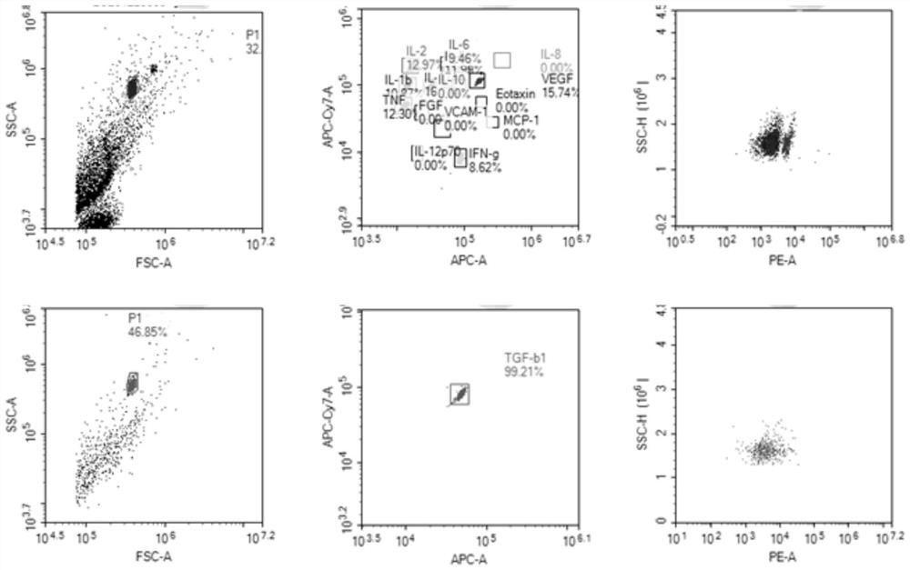
[0065] A detection reagent for intraocular fluid cytokines used for auxiliary diagnosis of inflammatory eye diseases, using HumanEotaxin Flex Set, Human IL-10 Flex Set, Human IL-12p70 Flex Set, Human IL-1βFlex Set, Human IL-6 Flex Set , Human IL-8 Flex Set, Human MCP-1 Flex Set, HumanTNF Flex Set, Human IFN-γ Flex Set and Human VEGF Flex Set detection reagents.
[0066] The detection method comprises the following steps:
[0067] Intraocular fluid sample preparation: take the vitreous humor and centrifuge it at 10000rpm for 10min to obtain the intraocular fluid sample;
[0068] Preparation of microsphere solution: Take 0.25 μl of each microsphere reagent and mix, add 10 μl of microsphere diluent, and mix well to obtain a microsphere solution;
[0069] Preparation of antibody solution: Take 0.25 μl of each antibody reagent and mix, add 10 μl of antibody diluent, mix well to obtain antibody solution;
[0070] Sample incubation: put the microsphere solution into a blank flow tu...
Embodiment 2
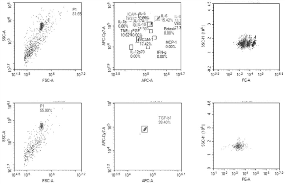
[0075] A method for detecting intraocular fluid cytokines for auxiliary diagnosis of immune eye diseases, using Human IL-1β Flex Set, Human IL-2 Flex Set, Human IL-5 Flex Set, Human IL-6 Flex Set, HumanTNF Flex Set , Human IFN-γ Flex Set, Human VEGF Flex Set, Human IL-17a Flex Set and Human TGF-β1 Single Plex Flex Set detection reagents.
[0076] The method for detecting cytokines other than TGF-β1 includes the following steps:
[0077] Intraocular fluid sample preparation: take aqueous humor and centrifuge at 10000rpm for 10min to obtain intraocular fluid sample;
[0078] Preparation of microsphere solution: Take 0.25 μl of each microsphere reagent except TGF-β1 and mix them, add 10.5 μl of microsphere diluent, and mix well to obtain a microsphere solution;
[0079] Antibody solution preparation: Take 0.25 μl of each antibody reagent except TGF-β1 and mix, add 10.5 μl of antibody diluent, mix well to obtain antibody solution;
[0080] Sample incubation: put the microsphere ...
Embodiment 3
[0094] A method for detecting intraocular fluid cytokines for auxiliary diagnosis of vascular eye disease, using Human IL-10 Flex Set, Human IL-6 Flex Set, Human IL-8 Flex Set, Human TNF Flex Set, HumanVEGF Flex Set, Human Soluble CD54 (ICAM-1) Flex Set, Human Soluble CD106 (VCAM-1) Flex Set, Human TGF-β1 Single Plex Flex Set detection reagents.
[0095] The method for detecting cytokines other than TGF-β1 includes the following steps:
[0096] Intraocular fluid sample preparation: take aqueous humor and centrifuge at 10000rpm for 10min to obtain intraocular fluid sample;
[0097] Preparation of microsphere solution: Take 0.25 μl of each microsphere reagent except TGF-β1 and mix them, add 10.75 μl of microsphere diluent, and mix well to obtain a microsphere solution;
[0098] Preparation of antibody solution: Take 0.25 μl of each antibody reagent except TGF-β1 and mix them, add 10.75 μl of antibody diluent, mix well to obtain antibody solution;
[0099] Sample incubation: pu...
PUM
 Abstract
Description
Claims
Application Information
 - R&D
- Intellectual Property
- Life Sciences
- Materials
- Tech Scout
- Unparalleled Data Quality
- Higher Quality Content
- 60% Fewer Hallucinations
Browse by: Latest US Patents, China's latest patents, Technical Efficacy Thesaurus, Application Domain, Technology Topic, Popular Technical Reports.
© 2025 PatSnap. All rights reserved.Legal|Privacy policy|Modern Slavery Act Transparency Statement|Sitemap|About US| Contact US: help@patsnap.com


