Lung medical image analysis method, device and system

A medical image and image analysis technology, which is applied in the fields of equipment and systems, lung medical image analysis, and lung lesion analysis, can solve the problems of impossible analysis of various lung diseases, and achieve high recognition and classification accuracy, The effect of improving computing efficiency and improving accuracy

Inactive Publication Date: 2021-07-09
SICHUAN UNIV
View PDF7 Cites 5 Cited by
  • Summary
  • Abstract
  • Description
  • Claims
  • Application Information

AI Technical Summary

Problems solved by technology

Further, the method of the above patent application is even more impossible...

Method used


Image

  • Lung medical image analysis method, device and system
  • Lung medical image analysis method, device and system
  • Lung medical image analysis method, device and system

Examples


Embodiment 1

[0043] Embodiment 1 A device for lung lesion analysis

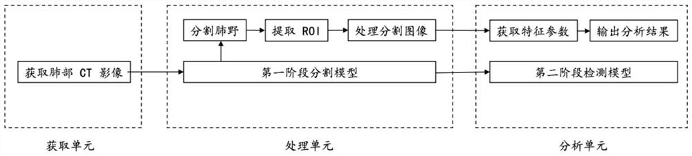
[0044] The device and the computer programs it runs such as figure 1 , 2 As shown, the device in this embodiment includes the following functional modules: an acquisition unit, a processing unit, and an analysis unit.

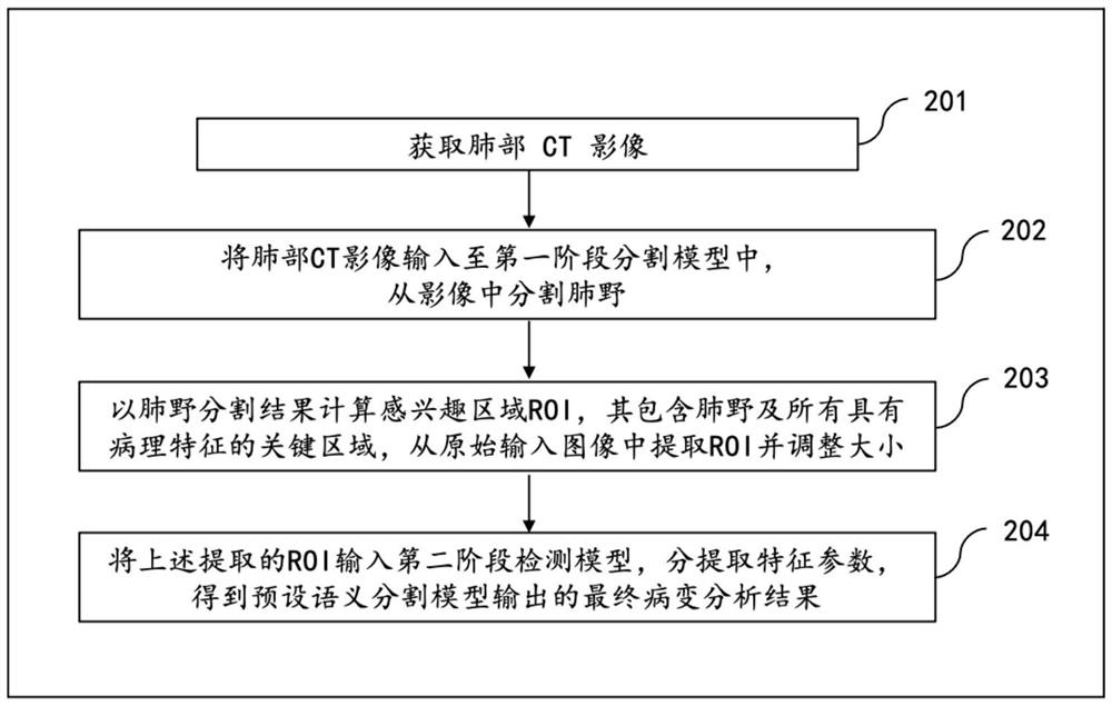
[0045] The acquiring unit is used for acquiring medical images of the patient's lungs. The medical images of the lungs are selected from computed tomography (Computed Tomography, CT for short) images and digital radiography (digital radiography, DR) images.

[0046] The processing unit is used to input the lung medical image into the first-stage segmentation model, segment the lung field, and use it as input, calculate the region of interest ROI with the lung field segmentation result, crop the ROI from the original input image and adjust the size , to get the input image for image recognition.

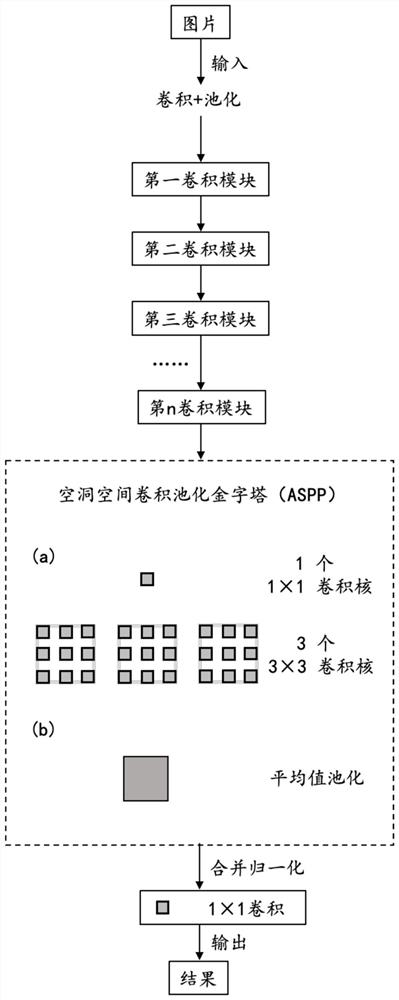
[0047]As a preferred solution, in the processing unit, the input medical image of the lungs is sampled at a...

Embodiment 2

[0055] Embodiment 2 A kind of computer device for lung lesion analysis

[0056] A computer device for the analysis of medical images of the lungs, structured as Figure 4 As shown, at least one processor 501 is included, and a storage 502 connected with the at least one processor. In this embodiment, the storage 502 has computer programs executable by at least one processor 501 .

[0057] Among them, the processor 501 is the calculation and control center of the lung lesion detection equipment, which is connected to various parts of the lung lesion detection equipment through various interfaces and lines, and can execute the instructions stored in the memory 502 or call the instructions stored in the memory The data in 502 is used to realize the function of lung lesion detection. In this embodiment, the processor 501 includes the acquisition unit, processing unit, and analysis unit described in Embodiment 1.

[0058] Optionally, the processor 501 includes one or more proces...


PUM


Abstract

The invention belongs to the technical field of machine learning, and particularly relates to a lung medical image analysis method, device and system. The lung medical image analysis method comprises the steps of segmenting a lung field and a lung lesion area from an acquired lung medical image, calculating an ROI (Region of Interest) containing the lung field and the lung lesion area, and cutting an input picture for image recognition in the lung medical image according to the ROI. And inputting the input picture into a second-stage detection model, further extracting feature parameters, and obtaining a final lesion analysis result output by a preset semantic segmentation model. By means of the lung medical image analysis method, device and system, multiple lung lesions can be recognized and classified and judged at the same time, and the device is high in efficiency and accuracy of lung medical image analysis and has good application prospects.

Description

technical field [0001] The invention belongs to the technical field of machine learning, and in particular relates to a lung medical image analysis method, equipment and system for analyzing lung lesions. Background technique [0002] In recent years, lung diseases have always shown a high outbreak rate. In the diagnosis of lung diseases, medical imaging diagnosis, including methods such as CT imaging diagnosis, plays an irreplaceable role. In the prior art, the CT diagnosis of the lungs is still performed by doctors manually observing the CT images and making judgments based on their own knowledge and experience, so as to find out the corresponding existing lesions. However, lung diseases have three characteristics in CT imaging manifestations: "different symptoms for different diseases, different symptoms for the same disease, and same symptoms for different diseases", which requires CT doctors to have extremely high theoretical knowledge and clinical experience. However...

Claims


Application Information

Patent Timeline
IPC IPC(8): G06T7/00G06K9/32G06K9/34G06K9/62G06N3/04
CPCG06T7/0012G06T2207/10081G06T2207/30061G06T2207/30096G06V10/25G06V10/267G06N3/045G06F18/214
Inventor 李经纬赵哲昊张琪然高子毅肖雨璇邱炳润翁义航奇鑫
Owner SICHUAN UNIV
Why Eureka
Try Eureka