Coronary artery plaque state evaluation method and device and electronic equipment

A coronary artery and state assessment technology, applied in the field of medical imaging, can solve the problems of ignoring lipid components, limited scanning depth, and inaccurate evaluation standards, and achieve the effects of improving positive predictive value, good evaluation effect, and improving reproducibility

Pending Publication Date: 2021-07-09
PULSE MEDICAL IMAGING TECH (SHANGHAI) CO LTD
View PDF0 Cites 2 Cited by
  • Summary
  • Abstract
  • Description
  • Claims
  • Application Information

AI Technical Summary

Problems solved by technology

Among them, although intravascular ultrasound (IVUS) and near-infrared spectroscopy (NIRS) technologies can assess the size of plaques in depth, the imaging mechanism itself still limits their ability to clearly distinguish and comprehensively evaluate the components of plaques.
[0004] Optical coherence tomography (OCT) has unique advantages in the evaluation of coronary artery plaques due to its high resolution, but due to its limited scanning depth, complete imaging of the entire plaque is still flawed
In addition, although OCT imaging technology can clearly display the thickness of the fibrous cap of coronary artery plaques, it cannot display the true size of the lipid components in the plaques due to optical attenuation.
At present, in clinical practice, based on OCT imaging technology, only the thickness of the fibrous cap <65um and the lipid angle ≥ 180° can be used as the evaluation criteria for unstable plaques, while the size of the lipid component, which has been proven to be important for stable plaques, is ignored. factors related to sexual status, the above evaluation criteria are not accurate enough

Method used


Image

  • Coronary artery plaque state evaluation method and device and electronic equipment
  • Coronary artery plaque state evaluation method and device and electronic equipment
  • Coronary artery plaque state evaluation method and device and electronic equipment

Examples


Embodiment 1

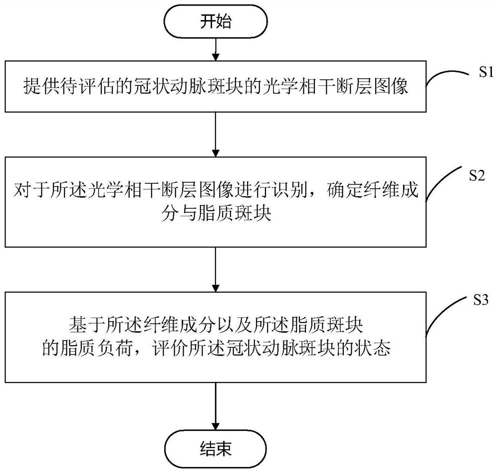
[0154] First, several frames of optical coherence tomography images of the coronary plaque to be evaluated are acquired. Figure 5 (a) shows 5 frames of optical coherence tomography images. Respectively, the original image of the target frame and the original images of the first before, the second before, the after 1, and the second after.

[0155] Next, for the 5 frames of the original image, the identification model is used for identification, and the identification result is as follows Figure 5 Shown in (b).

[0156] After identification, the fibrous components and lipid plaques were determined.

[0157] Next, based on the fiber components and lipid plaques in the identified images, the thickness of the fibrous cap and the lipid load in each frame were calculated.

[0158] The average value of the lipid load in the 5 frames of images and the median of the thickness of the fibrous cap were taken to calculate the LCR of the target frame.

[0159] Table 1 shows the calcul...


PUM


Abstract

The invention provides a coronary artery plaque state evaluation method and device, electronic equipment and a computer readable storage medium. The coronary artery plaque state evaluation method comprises the following steps: step S1, providing an optical coherence tomography image of a coronary artery plaque to be evaluated; S2, identifying the optical coherence tomography image, and determining fiber components and lipid plaques; and S3, evaluating the state of the coronary plaque based on the fiber component and the lipid load of the lipid plaque. According to the coronary artery plaque state evaluation method, the tomographic image of the blood vessel to be evaluated is obtained through the optical coherence tomography technology, the fiber components and the lipid plaque in the tomographic image are recognized based on the obtained tomographic image, and the state of the coronary artery plaque is comprehensively evaluated based on the recognized fiber components and lipid size. Quantitative evaluation can be realized, and the repeatability is higher.

Description

technical field [0001] The invention relates to the technical field of medical imaging, in particular to a method, device, electronic equipment and computer-readable storage medium for assessing the state of coronary artery plaque. Background technique [0002] Cardiac death is the leading cause of death in the population at present, and acute coronary syndrome caused by unstable plaque in coronary arteries is the main factor among them. Therefore, early detection of plaque stability in patients with coronary heart disease is crucial to the determination of treatment options and the assessment of long-term prognosis. [0003] Intracoronary imaging technology is currently the main means of assessing the stability of coronary plaques in patients with coronary heart disease. Among them, although intravascular ultrasound (IVUS) and near-infrared spectroscopy (NIRS) technologies can assess the size of plaques in depth, the imaging mechanism itself still limits their ability to c...

Claims


Application Information

Patent Timeline
IPC IPC(8): G06T7/00G06N3/04G06N3/08
CPCG06T7/0012G06N3/04G06N3/08G06T2207/10101G06T2207/20081G06T2207/30101
Inventor 常云霄李泽杭涂泽璇洪恢宏
Owner PULSE MEDICAL IMAGING TECH (SHANGHAI) CO LTD
Why Eureka
Try Eureka