Prostate cancer PDX model construction method
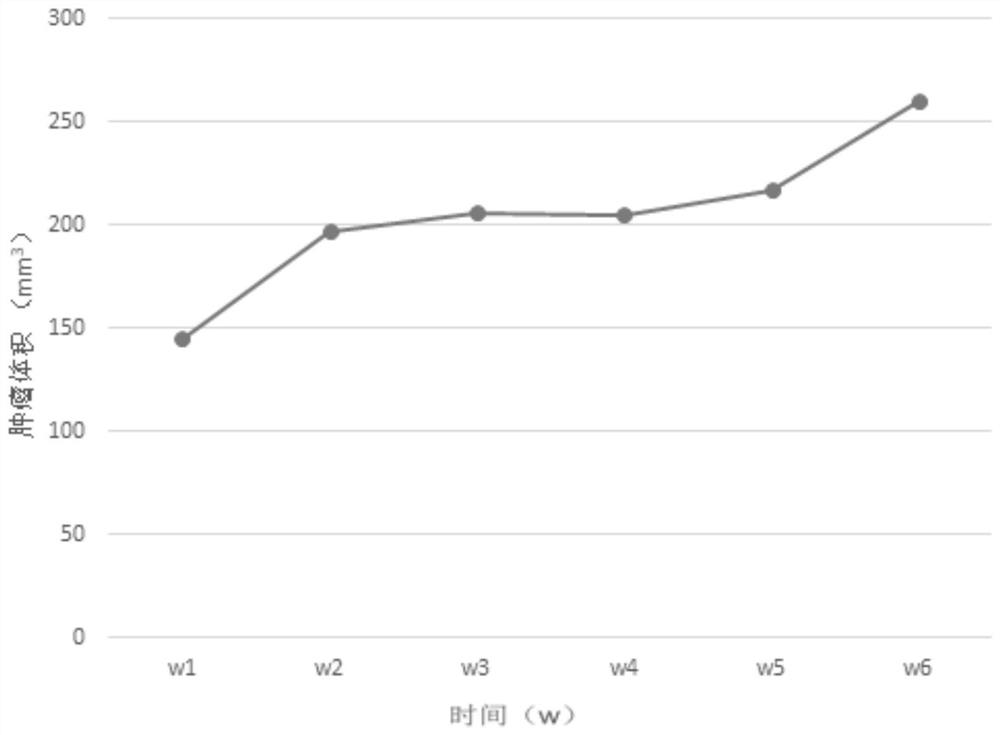
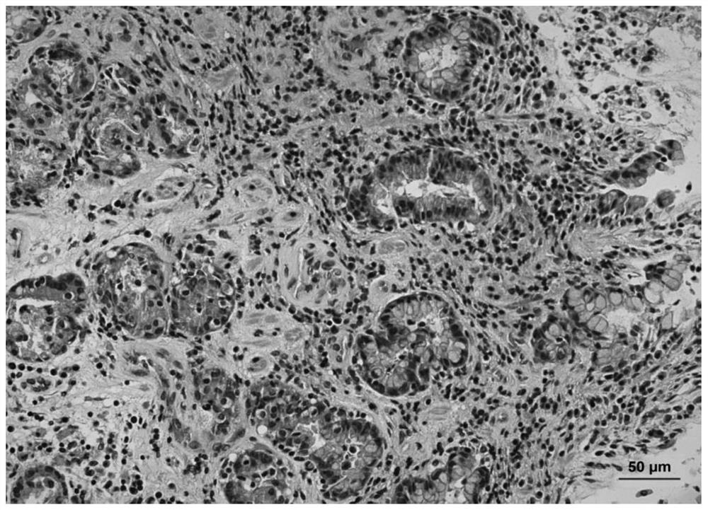
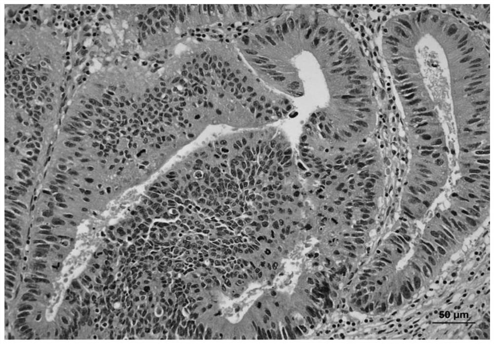
A prostate cancer and construction method technology, applied in the field of experimental animal models, can solve problems such as obstacles to clinical efficacy prediction and preclinical research, limited types, etc., and achieve the effect of overcoming limited animal models, low cost, and high tumor formation rate
- Summary
- Abstract
- Description
- Claims
- Application Information
AI Technical Summary
Problems solved by technology
Method used
Image
Examples
Embodiment 1
[0024] Example 1. A method for constructing a PDX model of prostate cancer
[0025] A method for constructing a prostate cancer PDX model, comprising the following steps:
[0026] 1) Cell extraction: Take a healthy B-NOD mouse, make a T-shaped incision on the lower abdomen, extract the seminal vesicles, peel off the superficial fascia, gently grind on a 200-mesh sieve, collect seminal vesicle cells, and mix them with a small amount of DMEM to obtain For seminal vesicle cell suspension, adjust the proportion of seminal vesicle cells in seminal vesicle cell suspension to 2×10 6 Individual / mL concentration;
[0027] 2) Resuscitate the animal tissue block and shred it to a size of 10-30mm 3 , mixing the shredded animal tissue block and seminal vesicle cell suspension at a volume ratio of 1:1 to obtain a tissue block containing seminal vesicle cells;
[0028] 3) Move the tissue block containing the seminal vesicle gland cells to a culture dish containing EPS to obtain a mixed so...
Embodiment 2
[0031] Example 2. A method for constructing a PDX model of prostate cancer
[0032] A method for constructing a prostate cancer PDX model, comprising the following steps:
[0033] 1) Cell extraction: Take a healthy B-NOD mouse, make a T-shaped incision on the lower abdomen, extract the seminal vesicles, peel off the superficial fascia, gently grind on a 200-mesh sieve, collect seminal vesicle cells, and mix them with a small amount of DMEM to obtain For seminal vesicle cell suspension, adjust the proportion of seminal vesicle cells in seminal vesicle cell suspension to 3×10 6 Individual / mL concentration;
[0034] 2) Resuscitate the animal tissue block and shred it to a size of 10-30mm 3 , mixing the shredded animal tissue block and seminal vesicle cell suspension at a volume ratio of 1:1 to obtain a tissue block containing seminal vesicle cells;
[0035] 3) Move the tissue block containing the seminal vesicle gland cells to a culture dish containing EPS to obtain a mixed so...
Embodiment 3
[0038] Example 3. A method for constructing a PDX model of prostate cancer
[0039] A method for constructing a prostate cancer PDX model, comprising the following steps:
[0040] 1) Cell extraction: Take a healthy B-NOD mouse, make a T-shaped incision on the lower abdomen, extract the seminal vesicles, peel off the superficial fascia, gently grind on a 200-mesh sieve, collect seminal vesicle cells, and mix them with a small amount of DMEM to obtain For seminal vesicle cell suspension, adjust the proportion of seminal vesicle cells in seminal vesicle cell suspension to 4×10 6 Individual / mL concentration;
[0041] 2) Resuscitate the animal tissue block and shred it to a size of 10-30mm 3 , mixing the shredded animal tissue block and seminal vesicle cell suspension at a volume ratio of 1:1 to obtain a tissue block containing seminal vesicle cells;
[0042] 3) Move the tissue block containing the seminal vesicle gland cells to a culture dish containing EPS to obtain a mixed so...
PUM
 Abstract
Description
Claims
Application Information
 - R&D
- Intellectual Property
- Life Sciences
- Materials
- Tech Scout
- Unparalleled Data Quality
- Higher Quality Content
- 60% Fewer Hallucinations
Browse by: Latest US Patents, China's latest patents, Technical Efficacy Thesaurus, Application Domain, Technology Topic, Popular Technical Reports.
© 2025 PatSnap. All rights reserved.Legal|Privacy policy|Modern Slavery Act Transparency Statement|Sitemap|About US| Contact US: help@patsnap.com


