
Dilator for vaginal endoscopic surgery of young women

A dilator and vaginal technology, applied in the field of medical devices, can solve the problems of patients' pain and discomfort, hymen injury, and inability to accurately locate, and achieve the effect of accurately positioning the scope of resected tissue, reducing the probability of injury, and promoting the outflow of hemorrhage

Pending Publication Date: 2021-08-13
THE THIRD AFFILIATED HOSPITAL OF GUANGZHOU MEDICAL UNIVERSITY
View PDF0 Cites 0 Cited by
  • Summary
  • Abstract
  • Description
  • Claims
  • Application Information

AI Technical Summary

Problems solved by technology

During use, the two leaves of the vaginal dilator stretch the labia and vagina, exposing the cervix and the vaginal fornix around it. The existing speculum has the following disadvantages: For young girls or unmarried women with asexual life history, place the speculum It will cause damage to the hymen and bring certain physical and psychological trauma, so this method is not suitable; there is no scale display, and the length cannot be accurately measured
As a result, it is impossible to accurately locate the surgical site, especially the tissue site with no obvious abnormality in appearance; the volume diameter is relatively large, the patient is prone to pain and discomfort, and even causes the patient's vagus nerve reflex stimulation; the two-leaf structure cannot provide all-round support for the vaginal wall, For patients with loose vaginal wall and large area or patients with oblique vaginal septum syndrome who have huge vaginal mass, normal tissue damage is prone to occur during vaginal endoscopic surgery

Method used


Image

  • Dilator for vaginal endoscopic surgery of young women
  • Dilator for vaginal endoscopic surgery of young women
  • Dilator for vaginal endoscopic surgery of young women

Examples


Embodiment 1

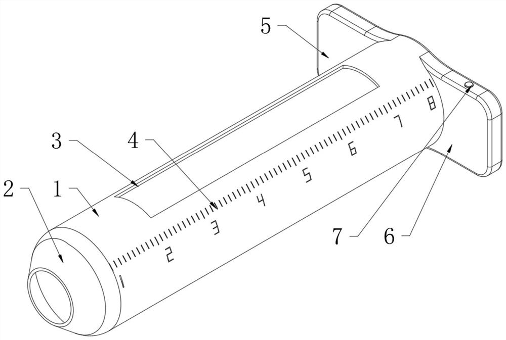
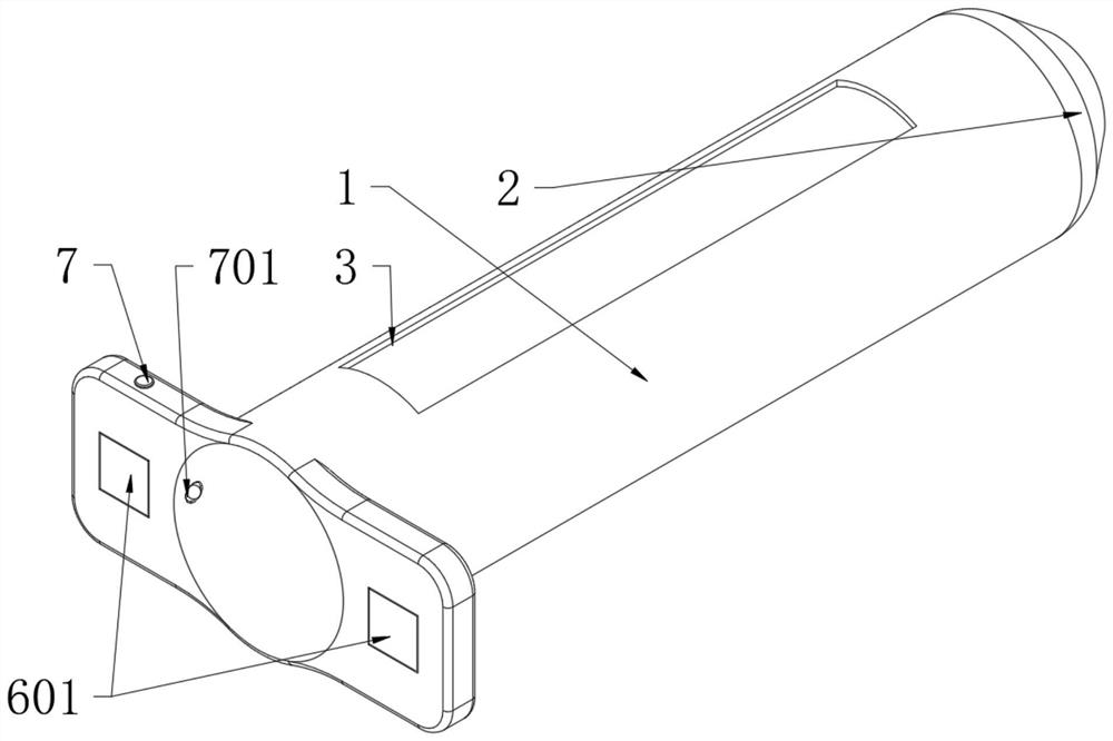
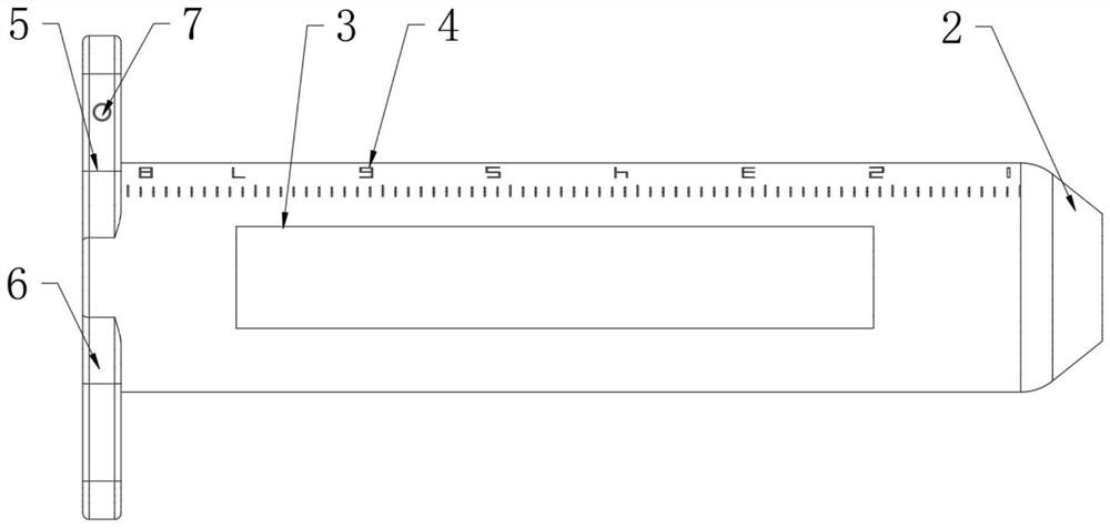
[0039] Such as Figure 1-5 As shown, a dilator for vaginal endoscopic surgery for young girls includes a dilator body 1, an opening is set at one end of the dilator body 1, a hand-held module is installed at the end of the dilator body 1, and a lighting mechanism is installed inside the dilator body 1; the opening is located at The end of the dilator main body 1 is a blunt eaves-shaped semi-opening 2; the hand-held module includes a first hand-held wing 5 and a second hand-held wing 6, and the first hand-held wing 5 and the second hand-held wing 6 are located away from the opening of the dilator main body 1 one end of which is fixed; the opening is a side window 3 and is embedded and installed on one side of the dilator main body 1; a scale line 4 is arranged on the outer wall of the dilator main body 1 and below the side window 3, the first hand-held wing 5 and the second The sides of the two hand-held wings 6 are embedded with magnetic blocks 601, and the magnetic blocks 601...


PUM


Abstract

The invention discloses a dilator for vaginal endoscopic surgery of young women. The dilator comprises a dilator body, an opening is formed in one end of the dilator body, a handheld module is installed at the end of the dilator body, and a lighting mechanism is installed on the inner side of the dilator body. The diameter of the dilator is smaller than that of a traditional vaginal speculum, the intact hymen can be effectively protected, in addition, the normal vaginal wall can be protected through one-side windowing, and the damage probability of non-surgical part vaginal wall tissue is reduced. A blunt brim-shaped semi-opening is formed in one end. A dome blunt brim is convenient for placement of the vagina and protection of the vaulted part of the vagina, and a central opening is convenient for observation of the cervix uteri. In order to solve the problems of huge oblique vaginal septum mass, large tension, poor vaginal distension effect of vaginal endoscopy or hysteroscope liquid and limited operation field, the rigid structure can effectively support the operation field, promote effusion of accumulated blood in the oblique vaginal septum mass, shorten the operation time and mark scales, so that the lengths of markers such as cervix uteri and hymen margins are visible, and the tissue excision range can be accurately positioned.

Description

technical field [0001] The invention relates to the technical field of medical instruments, in particular to an instrument suitable for endoscopic surgery on oblique vaginal septum syndrome of young girls. Background technique [0002] Existing vaginal dilators are used for voyeuristic examination, hysteroscopic surgery, vaginal surgery, and the like. During use, the two leaves of the vaginal dilator stretch the labia and vagina, exposing the cervix and the vaginal fornix around it. The existing speculum has the following disadvantages: For young girls or unmarried women with asexual life history, place the speculum It will cause damage to the hymen and bring certain physical and psychological trauma, so this method is not suitable; the lack of scale display makes it impossible to accurately measure the length. As a result, it is impossible to accurately locate the surgical site, especially the tissue site with no obvious abnormality in appearance; the volume diameter is re...

Claims


Application Information

Patent Timeline
Patent Type & Authority Applications(China)
IPC IPC(8): A61B17/02A61B17/42A61B18/12A61B90/00
CPCA61B17/0218A61B17/42A61B18/12A61B2018/00559A61B2018/00577A61B90/06A61B2090/061
Inventor 李竞雄谭鹰
Owner THE THIRD AFFILIATED HOSPITAL OF GUANGZHOU MEDICAL UNIVERSITY
Learn More