
Mounting unit and mounting method of analyte detection device

A technology for installing a unit and a detection device, applied in the field of medical devices, can solve problems such as poor reliability, signal loss, and inability to obtain analyte parameter data, and achieve the effects of avoiding breakage, improving reliability, and enhancing user experience.

Pending Publication Date: 2021-08-20
MEDTRUM TECH
View PDF22 Cites 2 Cited by
  • Summary
  • Abstract
  • Description
  • Claims
  • Application Information

AI Technical Summary

Problems solved by technology

However, the current detection device is prone to signal loss after installation, cannot obtain analyte parameter data, and has poor reliability

Method used


Image

  • Mounting unit and mounting method of analyte detection device
  • Mounting unit and mounting method of analyte detection device
  • Mounting unit and mounting method of analyte detection device

Examples


Embodiment Construction

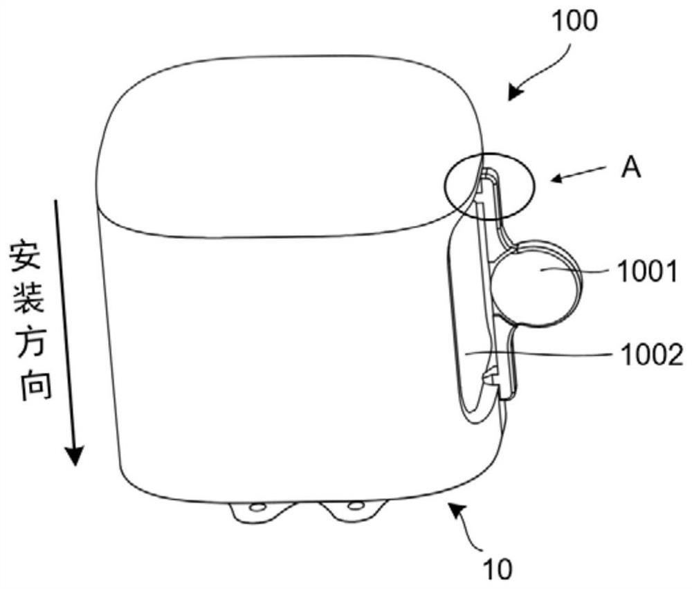
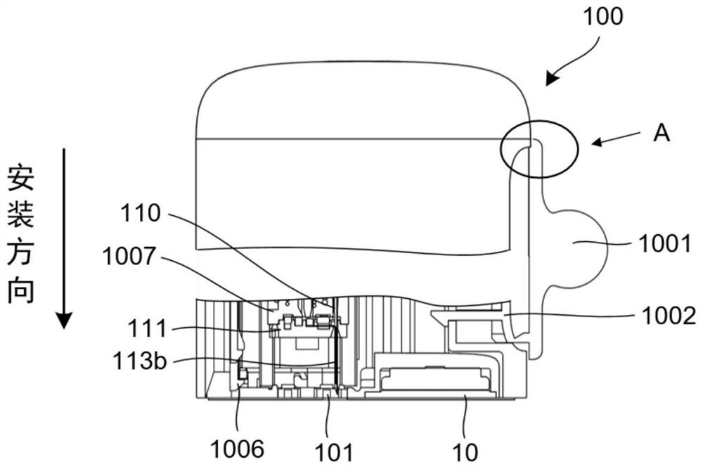
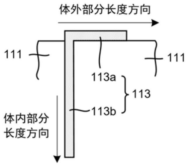
[0042] As mentioned above, after installation, the body fluid parameter detection device in the prior art is prone to failure, cannot obtain detection signals, and has poor reliability.

[0043] After research, it is found that the reason for the above problems is that the shape of the sensor needs to be changed during or after the installation of the existing detection device, which causes the electrodes or electrode leads on the surface of the sensor to break, causing signal interruption.

[0044] In order to solve this problem, the present invention provides an installation unit and installation method of an analyte detection device. The shape of the sensor does not change from the beginning to the end, and the electrodes or electrode leads on the surface of the sensor are avoided from breaking, ensuring normal detection and improving the analyte detection device. reliability.

[0045] Various exemplary embodiments of the present invention will now be described in detail wi...


PUM


Abstract

The invention discloses a mounting unit of an analyte detection device. The mounting unit comprises an auxiliary mounting structure, a sensor unit, a triggering part and a base, wherein the auxiliary mounting structure is arranged at the rear end of the mounting unit; the sensor unit is arranged close to the front end of the mounting unit; the sensor unit comprises a pedestal and a sensor arranged on the pedestal; the sensor comprises an in-vivo part and an in-vitro part; the in-vitro part is bent or buckled towards the top of the pedestal, and the in-vitro part is located at a working position; before the triggering part is started, the auxiliary mounting structure does not implement the mounting action; and the base is embedded into the mounting unit from the front end of the mounting unit. In the whole installation process, the shape of the in-vitro part of the sensor is not changed, an electrode or an electrode lead on the surface of the sensor is prevented from being broken, normal detection is ensured, and the reliability of the analyte detection device is improved.

Description

technical field [0001] The invention mainly relates to the field of medical devices, in particular to an installation unit of an analyte detection device and an installation method thereof. Background technique [0002] The pancreas in a normal human body can automatically monitor the glucose content in the human blood and automatically secrete the required insulin / glucagon. The function of the pancreas in diabetic patients is abnormal and cannot normally secrete the insulin needed by the human body. Therefore, diabetes is a metabolic disease caused by abnormal pancreatic function, and diabetes is a lifelong disease. At present, medical technology is still unable to cure diabetes, and the occurrence and development of diabetes and its complications can only be controlled by stabilizing blood sugar. [0003] Diabetics need to check their blood sugar before injecting insulin into their body. At present, most detection methods can continuously detect blood glucose, and send ...

Claims


Application Information

Patent Timeline
Patent Type & Authority Applications(China)
IPC IPC(8): A61B5/1473A61B5/00
CPCA61B5/14532A61B5/1473A61B5/14503A61B5/6801A61B5/6849A61B2560/063
Inventor 杨翠军
Owner MEDTRUM TECH
Learn More