Nuclear medicine multi-mode lesion image detection method and system

A technology of image detection and nuclear medicine, applied in the field of medical image processing, can solve problems such as complex operation methods, and achieve the effect of simple principle and strong compatibility

Inactive Publication Date: 2021-09-07
ATOMICAL MEDICAL EQUIP FO SHAN LTD +1
View PDF2 Cites 0 Cited by
  • Summary
  • Abstract
  • Description
  • Claims
  • Application Information

AI Technical Summary

Problems solved by technology

However, the imaging characteristics and data sizes of different devices and modalities are often inconsistent. Conventional methods usually rely on modal preprocessing such as scale change, registration, etc. to unify the modal data, and the operation method is too complicated.

Method used


Image

  • Nuclear medicine multi-mode lesion image detection method and system
  • Nuclear medicine multi-mode lesion image detection method and system
  • Nuclear medicine multi-mode lesion image detection method and system

Examples


Embodiment Construction

[0028] The technical solutions of the present invention will be further described below in conjunction with the accompanying drawings and through specific implementation methods.

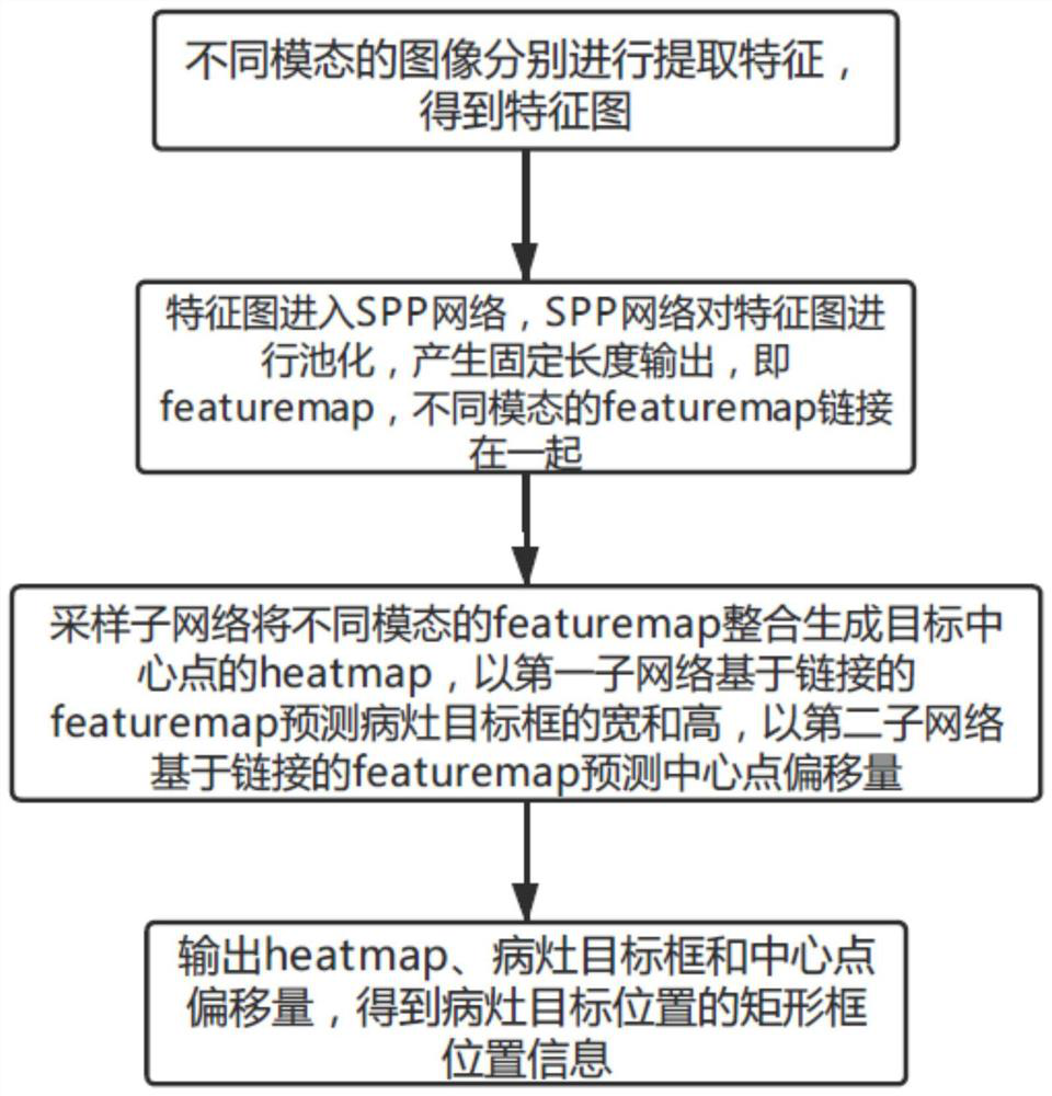
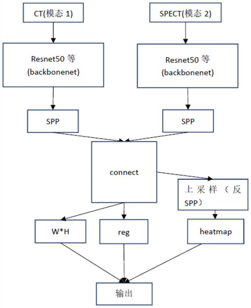
[0029] An example of this application, such as figure 1 As shown, a nuclear medicine multi-modal lesion image detection method is applied to nuclear medicine-based imaging equipment, and the nuclear medicine-based imaging equipment can be multi-modal imaging equipment, or can be of different types A single modality imaging machine, which comprises the following steps:

[0030] The features of images of different modalities are extracted separately to obtain feature maps.

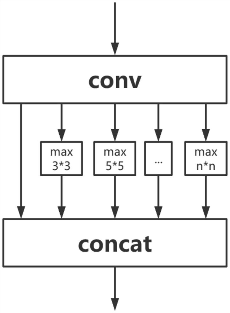
[0031] The feature map enters the SPP network, and the SPP network pools the feature map to generate a fixed-length output, that is, the featuremap, and the featuremaps of different modalities are linked together.

[0032] The sampling subnetwork integrates featuremaps of different modalities to generate a heatmap of the target ...


PUM


Abstract

The invention discloses a nuclear medicine multi-mode lesion image detection method and system. The method and system can effectively carry out the detection and integration processing of a lesion target image through the information of different modes, do not need extra multi-mode preprocessing work, and can comprehensively and completely reserve the information of each mode. After multi-scale feature fusion, the information of different modes is integrated, detail information of the different modes can be effectively reserved, and subsequent target detection by using the information of the different modes is facilitated. The principle part of a target detection head adopts the Centernet design concept in the prior art, a focus target is described as a central point, other characteristics such as the size of the target are obtained through direct regression in a feature map, and thus, the method is simple in principle and high in compatibility, does not need complex post-processing and achieves end-to-end detection.

Description

technical field [0001] The invention relates to the field of medical image processing, in particular to a nuclear medicine multimodal focus image detection method and system. Background technique [0002] Most of the current medical lesion detection methods are limited to one modality, such as CT, PET, MRI, SPECT, etc. alone. Restricted by its imaging principle, a single modality is often not enough for accurate processing of lesion images and follow-up personnel's judgment of lesions. Integrate different medical imaging equipment such as CT, PET, MRI, SPECT or different molecular probes, etc., integrate the advantages of various molecular imaging technologies, and develop a multi-modal fusion molecular imaging technology that can simultaneously provide the anatomical structure level of biological processes The combination of different modal information of information at the level of metabolism, function, physiology and pathology, and molecular and cellular levels makes use...

Claims


Application Information

Patent Timeline
Patent Type & Authority Applications(China)
IPC IPC(8): G06T7/00G06T7/60G06T5/50
CPCG06T7/0012G06T7/60G06T5/50G06T2207/20221
Inventor 杨雪松刘豆豆邓晓其他发明人请求不公开姓名
Owner ATOMICAL MEDICAL EQUIP FO SHAN LTD
Why Eureka
Try Eureka