In vivo in situ induced CAR-T cell delivery system targeting tumor and its application

A delivery system and tumor technology, applied in the field of immuno-oncology, can solve problems not related to the treatment of solid tumors, avoid complex processes and high costs, and simplify operations

Active Publication Date: 2022-07-19
XIEHE HOSPITAL ATTACHED TO TONGJI MEDICAL COLLEGE HUAZHONG SCI & TECH UNIV
View PDF0 Cites 0 Cited by
  • Summary
  • Abstract
  • Description
  • Claims
  • Application Information

AI Technical Summary

Problems solved by technology

It can be seen that the way NPs deliver CAR genes has good application prospects, but it has not yet been involved in the treatment of solid tumors

Method used


Image

  • In vivo in situ induced CAR-T cell delivery system targeting tumor and its application
  • In vivo in situ induced CAR-T cell delivery system targeting tumor and its application
  • In vivo in situ induced CAR-T cell delivery system targeting tumor and its application

Examples


Embodiment 1

[0039] Example 1 Delivery system for in situ induction of CAR-T cells targeting tumors in vivo

[0040] 1. Preparation of PBAE / pDNA

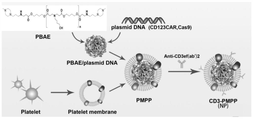
[0041] Dilute the PBAE and CD123 gene and CRISPR plasmids (purchased by the company) in 25mM sodium acetate solution respectively, add the PBAE solution dropwise to the same volume of CRISPR plasmid solution and mix, then add the CD123 gene plasmid and mix well. Assembly to construct PBAE / pDNA nanoparticles (e.g. figure 1 shown), wherein the plasmid ratio of CD123 gene and CRISPR is 1:1, and the ratio of PBAE to CD123 gene plasmid is 30:1.

[0042] 2. Preparation of platelet membrane-encapsulated PBAE / pDNA nanocarriers

[0043] After C57BL / 6J mice were anesthetized, blood was collected from the living heart in an anticoagulant tube, platelet separation solution was added, centrifuged at 300 g for 15 min, the first plasma layer was taken into a sterile centrifuge tube, and the same volume of sample diluent was added. , centrifuged at 500g for ...

Embodiment 2

[0045] Example 2 Characterization and identification of nanoparticles

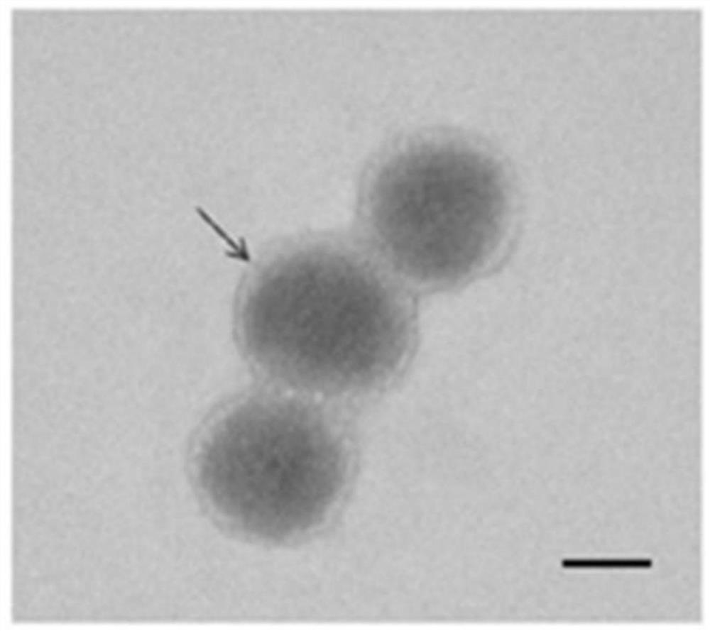
[0046] The morphology of the nanocarrier prepared in Example 1 was observed by transmission electron microscope, 5ul of the nanocarrier was diluted with deionized water 3 times, and then a small amount of the diluted sample was drawn and added dropwise to the copper mesh. The results are as follows: figure 2 As shown, the particle size of nanometers is 150-200 nm.

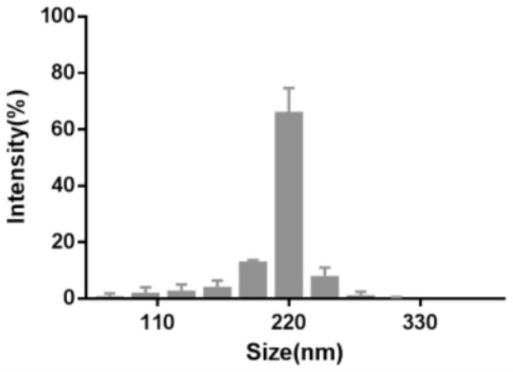
[0047] The size and zeta potential of nanoparticles were measured by dynamic light scattering laser nanoparticle size analyzer, and 1 mL of nanocarrier samples were taken into the particle size analyzer sample cell and the potential measurement sample cell, respectively. The results are as follows image 3 and Figure 4 As shown, the main particle size distribution of the nanoparticles is at 220 nm, and the zeta potential of the final encapsulated nanoparticle is -18 mV.

Embodiment 3

[0048] Example 3 Efficiency detection of nanoparticle transfection of plasmid into T cells in vitro

[0049] Pure pDNA, PBAE / pDNA and platelet membrane-encapsulated PBAE / pDNA groups were cultured with mouse spleen-derived T cells (PBMC), and the transfection efficiency was observed by confocal microscopy at 20 and 120 min of transfection. The result is as Figure 5 As shown, the platelet membrane-encapsulated PBAE / pDNA nanocarriers have good transfection efficiency compared to the plasmid pDNA group alone.


PUM

PropertyMeasurementUnit
particle diameteraaaaaaaaaa

Abstract

The invention discloses a delivery system for targeting tumors in vivo in situ inducing CAR-T cells and applications thereof, belonging to the technical field of immuno-oncology. The delivery system includes nanoparticles and a nanocarrier formed by wrapping the nanoparticles with a platelet membrane; wherein, the nanoparticles are assembled from a cationic polymer, a CAR gene plasmid and a CRISPR system, and the CRISPR is delivered through the nanocarrier The system enables in situ editing of T cells into the tumor. The present invention realizes in situ editing of T cells in solid tumors, improves the tumor microenvironment, and enhances the proliferation and persistence of CAR-T cells. Compared with traditional CAR-T, the invention has higher safety, simple process and low cost.

Description

technical field [0001] The present invention relates to the technical field of immuno-oncology, in particular to a delivery system for targeting tumors in vivo in situ inducing CAR-T cells and applications thereof. Background technique [0002] In recent years, CAR-T cell immunotherapy (ie, chimeric antigen receptor T cell immunotherapy) has shown good targeting, lethality and durability in clinical trials, and has made breakthroughs in the treatment of hematological tumors, becoming the research hotspot. With the approval of Novartis' Kymrial by the US FDA in August 2017, it marks that CAR-T cell therapy has truly entered clinical application. However, CAR-T therapy has poor therapeutic effect in solid tumors such as lymphoma, mainly due to: 1) The lack of specific antigens on the surface of tumor cells. Solid tumors lack a tumor-associated antigen similar to CD19 that exists specifically in blood tumors, resulting in the molecular targets of CAR-T cells appearing on the ...

Claims


Application Information

Patent Timeline
Patent Type & Authority Patents(China)
IPC IPC(8): A61K39/00A61K48/00A61P35/00
CPCA61K48/0025A61K48/005A61K39/001129A61P35/00
Inventor 吴小艳胡振华
Owner XIEHE HOSPITAL ATTACHED TO TONGJI MEDICAL COLLEGE HUAZHONG SCI & TECH UNIV
Why Eureka
Try Eureka