

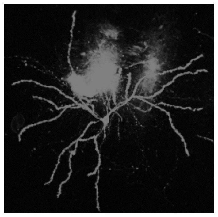
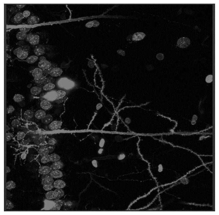
Method for detecting form of neuron dendritic spine in brain slice
A detection method and neuron technology, applied in the field of brain neuron research, can solve the problems of complex steps, long process, high cost, etc., and achieve the effect of simple operation steps, stable dyeing effect, and avoiding high background interference
- Summary
- Abstract
- Description
- Claims
- Application Information
AI Technical Summary
Problems solved by technology
Method used
Image

Examples
Embodiment 1
[0038] 1. Brain Slice Preparation
[0039] 1.1 Prepare a 1.5% paraformaldehyde-PBS solution: Dissolve 15 g of paraformaldehyde in 1 L of PBS solution (0.01 mol / L phosphate buffer, pH=7.4, the PBS mentioned below are all the same solution).
[0040] 1.2 After the mice were deeply anesthetized, the heart was perfused with PBS solution for 5 minutes, and then fixed with 1.5% paraformaldehyde-PBS solution for 20 minutes.
[0041] 1.3 Take out the complete brain from the cranial cavity, and immediately place it on a vibrating microtome to slice with a thickness of 300 μm. After slicing, take a clean six-hole plate and soak the brain slices in PBS solution for later use.
[0042] 2. DIL staining
[0043] 2.1 Prepare DIL stock solution: fully dilute the DIL solid with DMSO to a stock solution with a concentration of 20mmol / L, and store the stock solution at -20°C.
[0044] 2.2 Prepare DIL working solution: dilute 20mmol / L DIL stock solution in 1.5ml EP tube with PBS solution to 5μ...
PUM
| Property | Measurement | Unit |
|---|---|---|
| Concentration | aaaaa | aaaaa |
| Thickness | aaaaa | aaaaa |
Abstract
Description
Claims
Application Information