Method and device for collecting cerebral apoplexy tissue window evaluation value and storage medium

An evaluation value, stroke technology, applied in the field of medical images, can solve the problems of inability to accurately obtain the mismatch degree of DWI-FLAIR, difficult to identify, etc.

Pending Publication Date: 2021-10-22
沈阳先进医疗设备技术孵化中心有限公司
View PDF0 Cites 1 Cited by
  • Summary
  • Abstract
  • Description
  • Claims
  • Application Information

AI Technical Summary

Problems solved by technology

[0003] At present, doctors evaluate the degree of DWI-FLAIR mismatch in patients mainly by visually observing the high signal in the DWI (Diffusion Weighted Imaging, magnetic resonance diffusion weighted imaging) sequence, and whether the signal in the corresponding area on the T2 FLAIR (magnetic resonance imaging fluid attenuated inversion recovery) sequence is Abnormal, the method of naked eye observation is relatively easy to judge the obvious abnormal signal of T2 FLAIR sequence, but it is difficult to identify the inconspicuous high signal of T2 FLAIR sequence, and it is impossible to accurately obtain the degree of DWI-FLAIR mismatch

Method used


Image

  • Method and device for collecting cerebral apoplexy tissue window evaluation value and storage medium
  • Method and device for collecting cerebral apoplexy tissue window evaluation value and storage medium
  • Method and device for collecting cerebral apoplexy tissue window evaluation value and storage medium

Examples


Embodiment Construction

[0071] Hereinafter, the present application will be described in detail with reference to the drawings and embodiments. It should be noted that, in the case of no conflict, the embodiments in the present application and the features in the embodiments can be combined with each other.

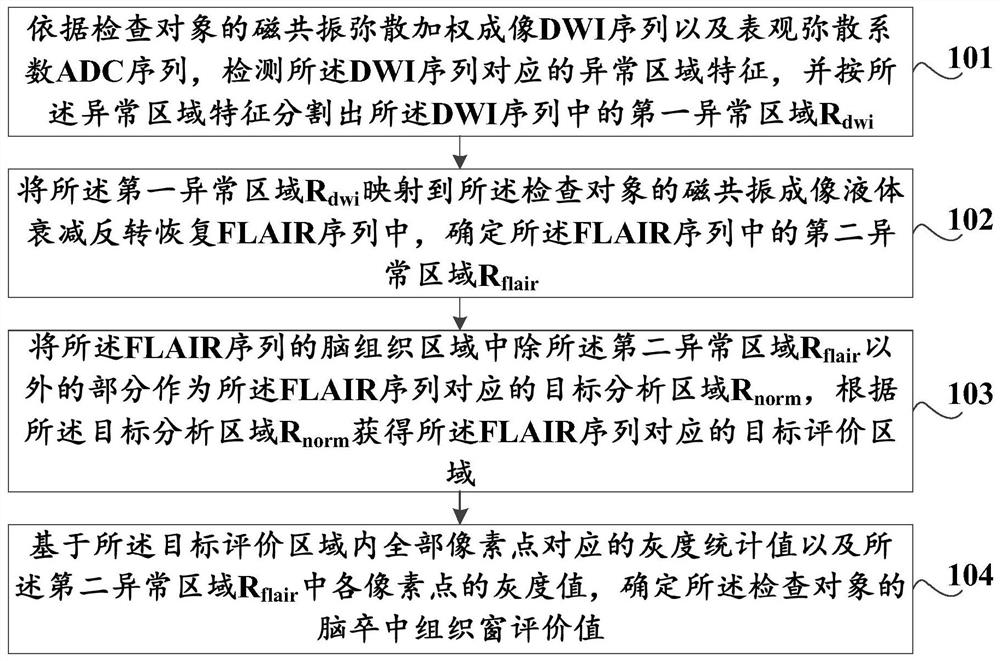
[0072] In this embodiment, a method for evaluating tissue windows in stroke is provided, such as figure 1 As shown, the method includes:

[0073] Step 101: According to the magnetic resonance diffusion weighted imaging DWI sequence and the apparent diffusion coefficient ADC sequence of the examination object, detect the characteristics of the abnormal region corresponding to the DWI sequence, and segment the first part of the DWI sequence according to the characteristics of the abnormal region. Abnormal area R dwi ;

[0074] Step 102, the first abnormal region R dwi Mapping to the magnetic resonance imaging fluid-attenuated inversion recovery FLAIR sequence of the examination object, and det...


PUM


Abstract

The invention discloses a method and a device for collecting a cerebral apoplexy tissue window evaluation value, a storage medium and computer equipment. The method comprises the following steps: detecting an abnormal region feature corresponding to a DWI sequence according to the DWI sequence and an ADC sequence, and segmenting a first abnormal region Rdwi in the DWI sequence; mapping the first abnormal region Rdwi to a FLAIR sequence of the inspection object, and determining a second abnormal region Rflag; taking the part, except the second abnormal region Rflag, in the brain tissue region of the FLAIR sequence as a target analysis region Rnorm, and obtaining a target evaluation region of the FLAIR sequence according to the target analysis region Rnorm; and determining a cerebral apoplexy tissue window evaluation value of the examination object based on the gray statistical values corresponding to all the pixel points in the target evaluation region and the gray values of all the pixel points in the second abnormal region Rflair.

Description

technical field [0001] The present application relates to the technical field of medical images, in particular to a method and device, a storage medium, and a computer device for obtaining an evaluation value of a stroke tissue window. Background technique [0002] Stroke is currently one of the major fatal and disabling diseases in all countries in the world. Stroke has become the first fatal disease in China and the leading cause of adult disability in China. For stroke patients, early intravenous thrombolysis and endovascular therapy can recanalize the occluded blood vessels and reperfuse the blood flow, thereby reducing the infarct volume, reducing the incidence of disability, and effectively improving the overall prognosis of patients. Therefore, how to make patients with acute cerebral infarction receive reperfusion therapy safely and effectively has become an urgent problem to be solved. The treatment of early stroke patients mainly depends on the time window, but wi...

Claims


Application Information

Patent Timeline
IPC IPC(8): G06T7/00G06T7/11G06T5/40G16H30/20
CPCG06T7/0012G06T5/40G06T7/11G16H30/20G06T2207/30016
Inventor 才品嘉李戈
Owner 沈阳先进医疗设备技术孵化中心有限公司
Why Eureka
Try Eureka