

Cell lysate and application thereof for ATP detection
A technology of cell lysate and lysis reaction, which is applied in the measurement/inspection of microorganisms, biochemical equipment and methods, etc. It can solve the problems of high storage requirements, repeated freezing and thawing, inconvenient use, etc., and achieves convenient operation, easy preparation, and stability sex-enhancing effect
- Summary
- Abstract
- Description
- Claims
- Application Information
AI Technical Summary
Problems solved by technology
Method used
Image

Examples
Embodiment 1
[0047] Embodiment 1: the selection of buffer system
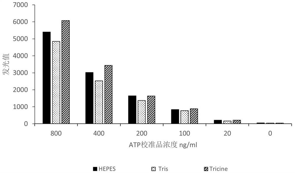
[0048] Select different types of buffer systems (HEPES, Tricine, Tris) commonly used for buffer system screening, and the reaction working solution includes the reaction solution (0.5%BSA, 40mm MgSO 4 , different buffers), luciferase (sigma), luciferin substrate (McLean), the reaction solution, luciferase, and luciferin substrate were mixed in a volume ratio of 1000:3:6, and different Concentration of ATP calibrator, 70ul / well, add 70ul of ATP reaction working solution to each well of ATP calibrator, keep it in the dark for 5min at 18-28°C, and use a chemiluminescence immunoassay analyzer to detect the ATP luminescence value.
[0049] Experimental results such as figure 1 shown. The results show that HEPES, Tricine and Tris buffer can all work, and the luminescence detection value is compared with Tris<HEPES<Tricine, and the Tricine buffer with higher detection value is preferred as the buffer in the lysate.
Embodiment 2
[0050]Example 2: Effects of different cell lysate compositions on ATP detection
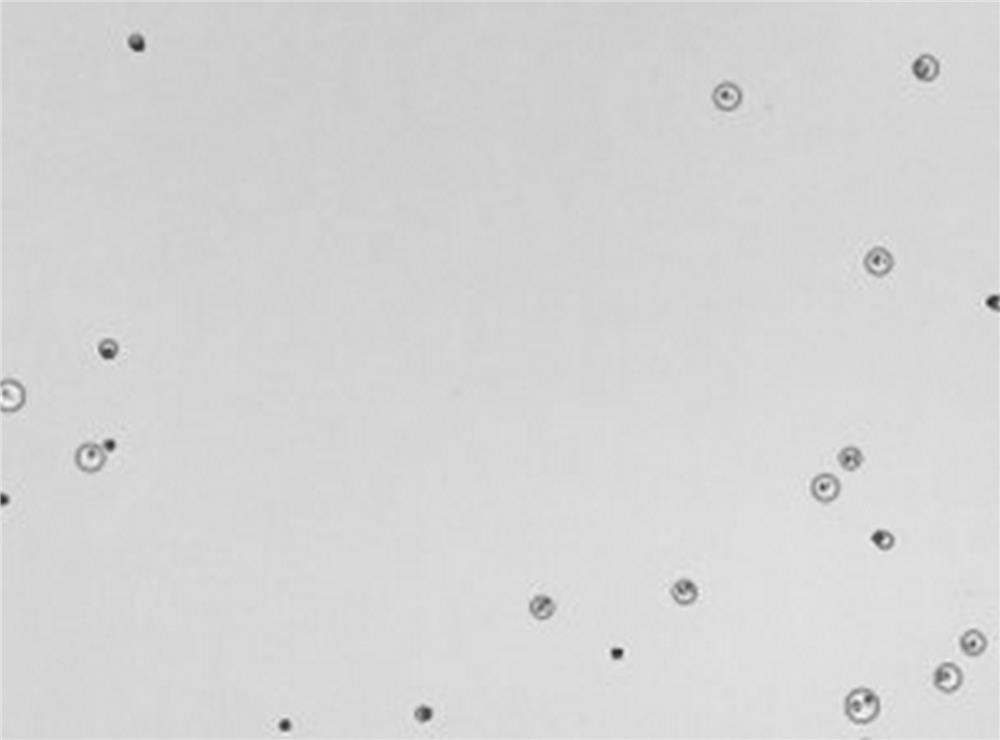
[0051] CD4 cells were magnetically sorted from PBMC cells (cell density was about 5×10 6 cells / ml), add 20ul CD4 cell suspension in the microwell, use 20mM Tricine buffer to configure the lysate according to Table 1, add 80ul lysate to the cell suspension and lyse for 10min, the magnetic frame absorbs the magnetic beads, take 70ul lysate to Add 70ul of ATP reaction working solution to the detection well, let it stand in the dark for 5 minutes at 18-28°C, and stabilize it for 10 minutes at room temperature. The ATP luminescence value is detected by chemiluminescence immunoassay. The detection results are shown in Table 2.
[0052] Table 1
[0053]
[0054] Table 2
[0055]
[0056] As can be seen from the results in Table 2, the use of lysis solutions containing NP-40, the luminescence values of different color development times are too low, and the ATP value after lysis can hardly be de...
Embodiment 3
[0057] Example 3: Effects of different compositions of cell lysate on ATP detection
[0058] CD4 cells were magnetically sorted from PBMC cells (cell density was about 5×10 6 cells / ml), add 20ul CD4 cell suspension (about 1×10 5 CD4 cells), add 80ul lysate to the cell suspension to lyse for 10min, absorb the magnetic beads on the magnetic frame, take 70ul lysate to the detection hole, add 70ul ATP reaction working solution, and stand in the dark for 5min at 18-28°C , stabilized at room temperature for 10 min, and used a chemiluminescence immunoassay analyzer to detect the luminescence value of ATP, and tested the influence of different compositions of cell lysates on the detection of ATP. The composition of the lysate 1-6 is shown in Table 3, and the test results are shown in Table 4.
[0059] table 3
[0060]
[0061] Table 4
[0062]
[0063] From the data in Table 4, it can be seen that the ATP detection value of lysate 1-5 can be basically stable above 90% within...
PUM
 Abstract
Description
Claims
Application Information