
Photoacoustic endoscopic device

A laser and optical fiber technology, applied in the field of endoscopy, can solve the problems of inability to achieve penetration depth and limited scanning range, and achieve the effects of large coverage, improved detection efficiency, and deep detection depth

Pending Publication Date: 2021-11-05
SHANGHAI NOVA OPTICS TECH
View PDF0 Cites 0 Cited by
  • Summary
  • Abstract
  • Description
  • Claims
  • Application Information

AI Technical Summary

Problems solved by technology

[0002] Photoacoustic endoscopic device is a device applied in the field of medical imaging, which can be used in subcutaneous tissue, rectum or prostate. Contrast, and can achieve a penetration depth that cannot be achieved by traditional optical imaging. It combines the advantages of the high contrast characteristics of pure optical imaging and the high penetration depth characteristics of pure ultrasound imaging, which improves the imaging clarity and avoids the influence of light scattering in principle. , broke through the "soft limit" of high-resolution optical imaging depth, and can realize deep molecular imaging in vivo. The current photoacoustic endoscopic device mainly uses circular scanning, and the scanning range of each point is limited, and requires a large scanning range, scanning A photoacoustic endoscopic probe with sufficient depth has certain shortcomings

Method used


Image

  • Photoacoustic endoscopic device
  • Photoacoustic endoscopic device
  • Photoacoustic endoscopic device

Examples


Embodiment Construction

[0044] The present invention will be described in detail below in conjunction with the accompanying drawings and specific embodiments. The present invention is not limited to this embodiment, and other embodiments may also belong to the scope of the present invention as long as they conform to the gist of the present invention.

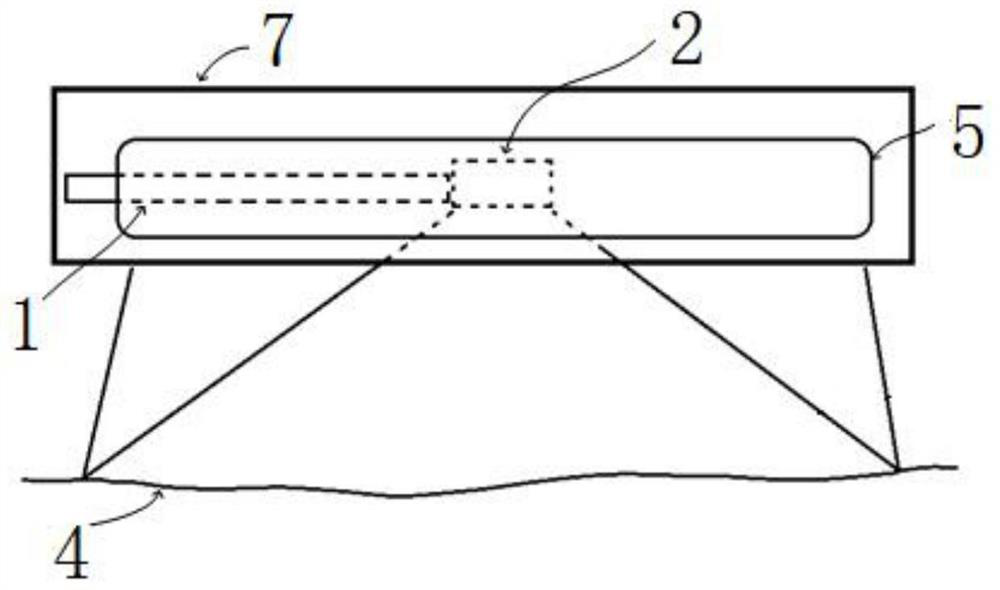
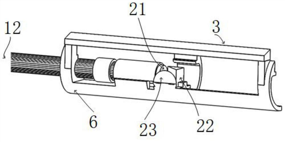
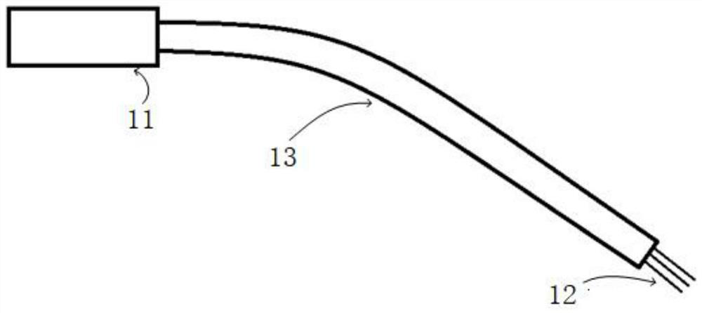
[0045] In a preferred embodiment of the present invention, based on the above-mentioned problems in the prior art, a photoacoustic endoscopic device is now provided, such as Figure 1-4 shown, including:

[0046] An optical fiber bundle 1 for conducting a laser;

[0047] An optical probe 2, connected to the optical fiber bundle 1, for coupling and shaping the laser light transmitted from the optical fiber bundle 1 and outputting it;

[0048] A long prism 3, arranged on one side of the optical probe 2, is used to converge the coupled and shaped laser output from the optical probe 2 and irradiate the laser light on a biological tissue 4;

[0049] An ...


PUM


Abstract

The invention provides a photoacoustic endoscopic device, and relates to the technical field of endoscopes. The photoacoustic endoscopic device comprises an optical fiber bundle, an optical probe, a long prism and an acoustic probe, wherein the optical fiber bundle is used for conducting laser; the optical probe is connected with the optical fiber bundle, and is used for coupling, shaping and outputting the laser conducted by the optical fiber bundle; the long prism is arranged at one side of the optical probe, and is used for gathering the coupled and shaped laser output by the optical probe and enabling the laser to irradiate a biological tissue; and the acoustic probe is arranged at one side, away from the optical probe, of the long prism, is confocal with an optical path of the optical probe, and is used for receiving ultrasonic waves generated by the laser irradiating the biological tissue, and obtaining a distribution image corresponding to the biological tissue according to the ultrasonic waves. The photoacoustic endoscopic device has the beneficial effects that the shaping of the laser beam from the optical fiber to the biological tissue is realized, the detection depth is deeper, and the detection width is larger, so that the larger coverage range is realized, and the medical endoscopic detection efficiency is improved.

Description

technical field [0001] The invention relates to the technical field of endoscopes, in particular to a photoacoustic endoscope device. Background technique [0002] Photoacoustic endoscopic device is a device used in the field of medical imaging, which can be used in subcutaneous tissue, rectum or prostate. Contrast, and can achieve a penetration depth that cannot be achieved by traditional optical imaging. It combines the advantages of the high contrast characteristics of pure optical imaging and the high penetration depth characteristics of pure ultrasound imaging, which improves the imaging clarity and avoids the influence of light scattering in principle. , broke through the "soft limit" of high-resolution optical imaging depth, and can realize deep molecular imaging in vivo. The current photoacoustic endoscopic device mainly uses circular scanning, and the scanning range of each point is limited, and it needs a large scanning range. A photoacoustic endoscopic probe with...

Claims


Application Information

Patent Timeline
IPC IPC(8): A61B5/00
CPCA61B5/0095
Inventor 杨波盛东波刘云鹏樊程祥王星琪赵倩阳
Owner SHANGHAI NOVA OPTICS TECH
Learn More