Multi-low-level feature fusion retinal vessel segmentation method

A technology of retinal blood vessels and low-level features, applied in the field of retinal blood vessel segmentation with multi-low-level feature fusion, can solve the problems of wasting computing power, limited segmentation effect, low efficiency, etc., and achieve the effect of high classification accuracy

Pending Publication Date: 2021-12-10
SOUTHWEST JIAOTONG UNIV
View PDF2 Cites 1 Cited by
  • Summary
  • Abstract
  • Description
  • Claims
  • Application Information

AI Technical Summary

Problems solved by technology

The main problems are as follows: 1. The image processing and feature extraction within the radius centered on the basic pixel point may lead to duplication or omission of the overall image processing, wasting computing power and low efficiency; 2. Multi-dimensional The selection of

Method used


Image

  • Multi-low-level feature fusion retinal vessel segmentation method
  • Multi-low-level feature fusion retinal vessel segmentation method
  • Multi-low-level feature fusion retinal vessel segmentation method

Examples


Embodiment 1

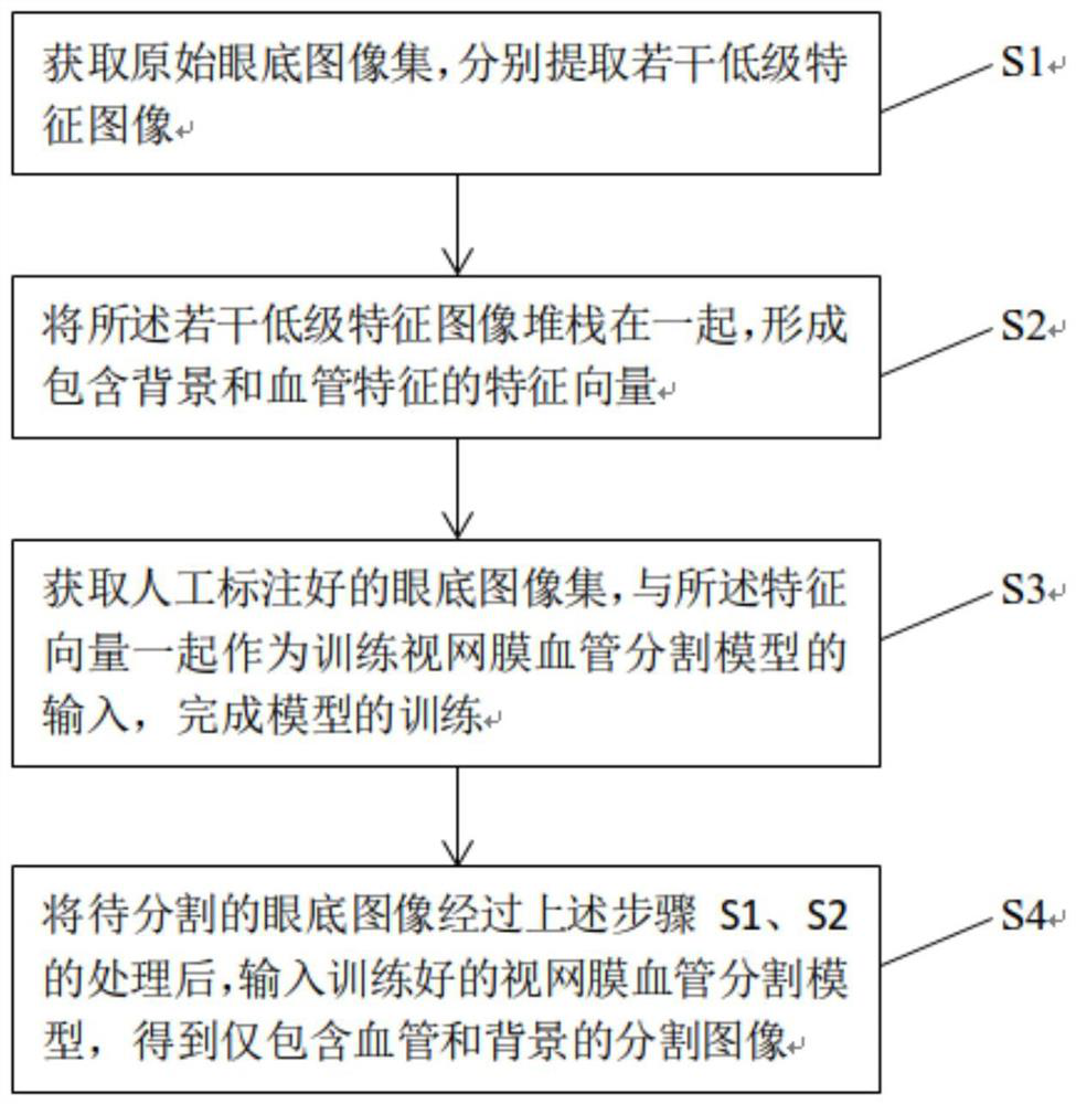
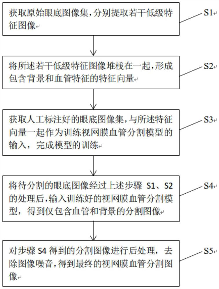
[0037] Such as figure 1 As shown, this embodiment provides a retinal blood vessel segmentation method with multi-low-level feature fusion, including steps:

[0038] Step S1. Obtain the original fundus image set, and extract several low-level feature images respectively;

[0039] Step S2. Stacking the several low-level feature images together to form a feature vector containing background and blood vessel features;

[0040] Step S3. Acquire a manually marked fundus image set, and use the feature vector as an input for training the retinal vessel segmentation model to complete the model training;

[0041] Step S4. After the fundus image to be segmented is processed in the above steps S1 and S2, it is input into the trained retinal blood vessel segmentation model to obtain a segmented image containing only blood vessels and background.

[0042] It should be understood that the fundus image contains various features, but not all features are helpful to improve the performance of...

Embodiment 2

[0045] This embodiment is developed on the basis of the above-mentioned embodiment 1. This embodiment provides a specific example of extracting low-level feature images, including: extracting several low-level feature images in step S1 includes extracting color feature images; Color profile images include:

[0046] Green channel feature map F in RGB color space Green ; red and orange mask F in HSV color space HSV .

[0047] It should be understood that the acquired fundus image is a color image, which includes red, green and blue channel features. For the retinal vessel image, the color fundus image shows the strongest contrast in the image separated by the green channel, so this embodiment selects the green feature map as an important low-level feature.

[0048] On the other hand, the HSV color space includes black, white, red, orange, yellow, green, cyan, blue, and purple, where, in the red space, the large blood vessels of fundus images are displayed more clearly, while ...

Embodiment 3

[0050] This embodiment is developed on the basis of the above-mentioned embodiment 1. This embodiment provides another specific example of extracting low-level feature images, including: extracting several low-level feature images as described in step S1 including the brightness image F Lum ; The brightness image F Lum The extraction formula is:

[0051] f Lum =0.299*R+0.587*G+0.114*B; wherein, R, G, and B are the red, green, and blue channel feature images of the fundus image in RGB color space, respectively. It should be understood that in the fundus image, blood vessels in each region have different brightness values ​​from the background, and different thresholds can effectively separate the blood vessels from the background. Therefore, this embodiment considers the brightness value as a feature. In the color retinal vascular images, the brightness of the blood vessels and the background in different regions are obviously different. Moreover, as described in the above-m...


PUM


Abstract

The invention provides a multi-low-level feature fusion retinal vessel segmentation method, which comprises the following steps of: S1, acquiring an original fundus image set, and respectively extracting a plurality of low-level feature images; s2, stacking a plurality of low-level feature images together to form a feature vector containing background and blood vessel features; s3, acquiring a manually labeled eye fundus image set, and taking the eye fundus image set and the feature vectors as input for training a retinal vessel segmentation model to complete model training; and S4, processing a fundus image to be segmented through the steps S1 and S2, and inputting the fundus image to be segmented into a trained retinal blood vessel segmentation model to obtain a segmented image only containing blood vessels and backgrounds. According to the method, multiple low-level features of the retinal blood vessel image features are fully considered, rich retinal blood vessel features can be effectively reserved, and the classification accuracy is high. The method does not depend on a large amount of sample data training, and solves the problems of few learning samples and poor effect of a deep learning method caused by the actual condition of small samples in the fundus image.

Description

technical field [0001] The invention relates to the technical field of medical imaging, in particular to a retinal blood vessel segmentation method based on fusion of multiple low-level features. Background technique [0002] Retinal blood vessels are part of human blood vessels. Many ophthalmic and cardiovascular diseases, such as glaucoma, diabetes, retinopathy, hypertension, arterial hardening etc. Computer-aided diagnosis (CAD) of fundus images could not only simplify mass screening of diabetic populations, but also allow clinicians to use time efficiently. Therefore, automatic detection and extraction of retinal vascular structures in color fundus images is of great significance. In fundus images, the retinal vascular network is intricate, and the brightness of vessels varies with the extension of vessels, making segmentation challenging. At present, there are mainly two types of blood vessel segmentation methods for fundus images: one is an unsupervised learning met...

Claims


Application Information

Patent Timeline
IPC IPC(8): G06T7/194G06T7/187G06K9/46G06K9/62
CPCG06T7/194G06T7/187G06T2207/30041G06T2207/30101G06F18/253
Inventor 邓涛黄怡张俊丰
Owner SOUTHWEST JIAOTONG UNIV
Why Eureka
Try Eureka