
Medical image analysis method and device based on plain-scan CT (Computed Tomography) data

A plain scan and data technology, applied in the field of medical image analysis, can solve the problems of low accuracy and large deviation of acute pulmonary embolism image analysis, and achieve the effect of improving accuracy

Inactive Publication Date: 2022-01-04
INFERVISION MEDICAL TECH CO LTD
View PDF9 Cites 6 Cited by
  • Summary
  • Abstract
  • Description
  • Claims
  • Application Information

AI Technical Summary

Problems solved by technology

However, this image analysis method based on plain scan CT data requires physicians to detect blood vessel diameter and shape changes by observing plain scan CT data, which is extremely related to physicians' clinical experience. low precision

Method used


Image

  • Medical image analysis method and device based on plain-scan CT (Computed Tomography) data
  • Medical image analysis method and device based on plain-scan CT (Computed Tomography) data
  • Medical image analysis method and device based on plain-scan CT (Computed Tomography) data

Examples


Embodiment Construction

[0055] In order to make the purpose, technical solutions and advantages of the embodiments of the present invention clearer, the technical solutions in the embodiments of the present invention will be clearly and completely described below in conjunction with the drawings in the embodiments of the present invention. Obviously, the described embodiments are only It is a part of embodiments of the present invention, but not all embodiments. The components of the embodiments of the invention generally described and illustrated in the figures herein may be arranged and designed in a variety of different configurations. Accordingly, the following detailed description of the embodiments of the invention provided in the accompanying drawings is not intended to limit the scope of the claimed invention, but merely represents selected embodiments of the invention. Based on the embodiments of the present invention, all other embodiments obtained by those skilled in the art without making...


PUM


Abstract

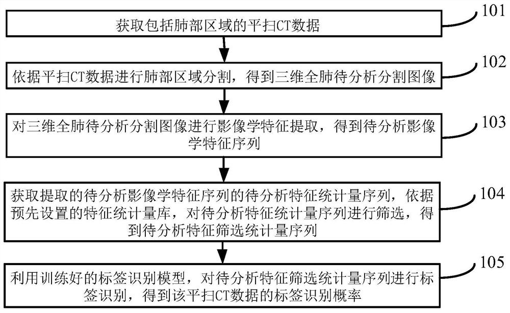
The invention provides a medical image analysis method and device based on plain-scan CT data. The method comprises the following steps: acquiring plain-scan CT data including a lung region; performing lung region segmentation according to plain-scan CT data, and obtaining a three-dimensional whole-lung image to be analyzed and segmented; performing imaging feature extraction on the three-dimensional whole-lung image to be analyzed and segmented to obtain a to-be-analyzed imaging feature sequence; obtaining a to-be-analyzed feature statistic sequence of the extracted to-be-analyzed iconography feature sequence, and screening the to-be-analyzed feature statistic sequence according to a preset feature statistic library to obtain a to-be-analyzed feature screening statistic sequence; and performing label identification on the to-be-analyzed feature screening statistic sequence by using the trained label identification model to obtain the label identification probability of the plain-scan CT data. The analysis precision of the acute pulmonary embolism on the plain-scan CT image can be improved.

Description

technical field [0001] The present invention relates to the technical field of image processing, in particular to a medical image analysis method and device based on unenhanced CT data. Background technique [0002] Pulmonary artery enhanced CT (CTPA, Computed Tomography Pulmonary Angiography) examination can show the filling defect of the pulmonary artery, so as to obtain the situation that the pulmonary artery is filled by thrombus, and it is an important examination method for image analysis of pulmonary embolism. In recent years, as the incidence of acute pulmonary embolism has increased year by year, it is very important to pre-analyze images of pulmonary embolism. [0003] At present, some patients cannot use contrast medium for pulmonary artery enhanced tomography (CT, Computed Tomography) due to some reasons, or, due to atypical cardiopulmonary symptoms, pulmonary artery enhanced CT cannot be performed in case patients, and only plain CT can be performed. The fillin...

Claims


Application Information

Patent Timeline
Patent Type & Authority Applications(China)
IPC IPC(8): G06T7/00G06T7/11
CPCG06T7/0012G06T7/11G06T2207/10081G06T2207/10028G06T2207/30061G06T2207/20081
Inventor 亢寒王少康陈宽
Owner INFERVISION MEDICAL TECH CO LTD
Learn More