Monoclonal antibody of novel coronavirus and mutant thereof and application of monoclonal antibody

A monoclonal antibody and antigen technology, applied in the fields of immunology and molecular virology, can solve problems affecting the effect of neutralizing antibodies

Active Publication Date: 2022-01-18
INST OF MICROBIOLOGY - CHINESE ACAD OF SCI
View PDF2 Cites 11 Cited by
  • Summary
  • Abstract
  • Description
  • Claims
  • Application Information

AI Technical Summary

Problems solved by technology

Studies have identified some viral mutations that can escape certain monoclonal antibodies, and these mutations occur at key positions where neutralizing antibodies bind to the virus, thereby affecting the effect of neutralizing antibodies

Method used


Image

  • Monoclonal antibody of novel coronavirus and mutant thereof and application of monoclonal antibody
  • Monoclonal antibody of novel coronavirus and mutant thereof and application of monoclonal antibody
  • Monoclonal antibody of novel coronavirus and mutant thereof and application of monoclonal antibody

Examples


Embodiment 1

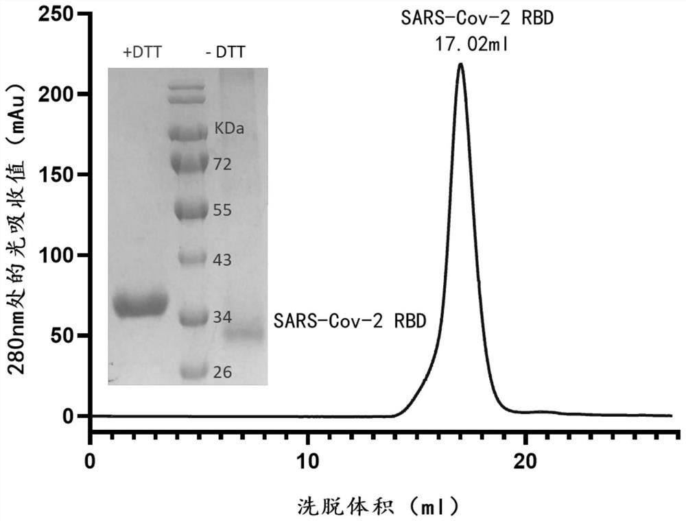
[0110] Example 1 Expression and purification of SARS-CoV-2 virus S protein RBD

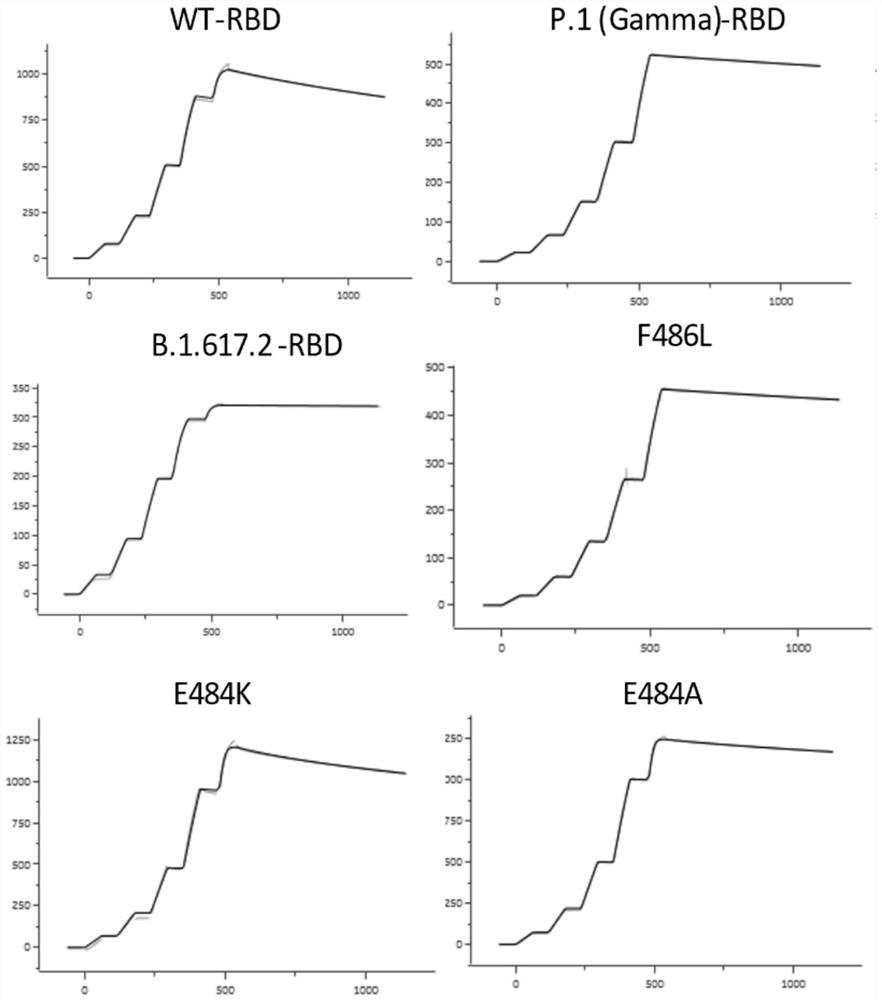
[0111] The optimized wild-type nCoV-RBD (residues 319-541, GenBank: YP_009724390.1) coding sequence with 6 His tags at the C-terminus was cloned into the mammalian expression vector pCAGGS. The various mutant RBDs shown in Table 1 (the mutation sites contained are as follows: K417N, K417T (present in P.1 (Gamma) RBD), L452R (present in B.1.617.1 (Kappa) RBD and In B.1.617.2 (Delta) RBD), Y453F, N460S, T478K (in B.1.617.2 (Delta) RBD), E484K, E484A, F486L, N501Y (in B.1.1.7 (Alpha) The coding sequences of RBD, B.1.351 (Beta) RBD, P.1 (Gamma) RBD), N501T) were subcloned into pCAGGS. Then the plasmid (2 μg) and PEI were transiently co-transfected at a mass ratio of 1:3 per milliliter of HEK293F cells. At 310K, 5% CO 2 Cells were cultured with SMM 293-TII medium (SinoBiological) under certain conditions, and then supplemented with SMS M293-SUPI (SinoBiological) at a ratio of 35 mL / L 24 hours after ...

Embodiment 2

[0112] Example 2 Isolation of memory B cells that specifically recognize RBD protein

[0113] With the informed consent of those infected with the SARS-CoV-2 virus who were cured and discharged, 10 mL of blood was collected and PBMCs were isolated. Separated PBMCs in 10 7 / mL density and the RBD protein prepared in Example 1 with a final concentration of 400nM were incubated on ice for half an hour; then washed twice with PBS, and then incubated with the following antibodies (both purchased from BD): anti-human CD3 / PE -Cy5, anti-human CD16 / PE-Cy5, anti-human CD235a / PE-Cy5, anti-human CD19 / APC-Cy7, anti-human CD27 / Pacific Blue, anti-human CD38 / APC, anti-human IgG / FITC, and anti-His / PE. After incubation on ice for half an hour, wash PBMCs twice with PBS. Subsequently, sort PBMCs with FACSAria III and collect PE - Cy5 - APCs - APCs - Cy7 + Pacific Blue + FITC + PE + The cells (that is, B cells) were directly collected into a 96-well plate, 1 cell / well.

Embodiment 324

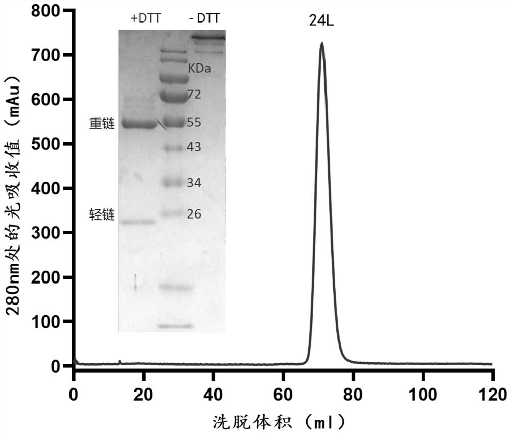
[0114] Example 3 Isolation and identification of 24L antibody and construction of recombinant expression vector

[0115] The B cells obtained in Example 2 were reverse-transcribed using Superscript III reverse transcriptase (Invitrogen) (at 55° C. for 60 minutes), wherein the reverse transcription primers used are shown in Table 2.

[0116] The sequence information of the reverse transcription primers used in Table 2

[0117]

[0118]Using the reverse transcription product as a template, the first round of PCR (PCRa) was carried out with HotStar Tap Plus enzyme (QIAgen) to amplify the sequence of the variable region of the antibody; wherein, the primers used are shown in Table 3; the reaction conditions used As follows: 95°C, 5min; 35 cycles of (95°C for 30s, 55°C (heavy chain / κ chain) for 30s, 72°C for 90s); 72°C, 7min. Subsequently, the second round of PCR (PCRb) was carried out using the amplified product as a template; wherein, the primers used were as shown in Table 4...


PUM


Abstract

The invention relates to the technical field of immunology and molecular virology, and particularly discloses a monoclonal antibody of novel coronavirus and a mutant thereof and application of the monoclonal antibody. Complementary determining regions CDR1, CDR2 and CDR3 of a heavy chain variable region of the monoclonal antibody or an antigen binding fragment thereof respectively have amino acid sequences as shown in SEQ ID NO: 1, SEQ ID NO: 2 and SEQ ID NO: 3; and complementary determining regions CDR1, CDR2 and CDR3 of a light chain variable region of the monoclonal antibody or the antigen binding fragment thereof respectively have amino acid sequences as shown in SEQ ID NO: 4, SEQ ID NO: 5 and SEQ ID NO: 6. The monoclonal antibody disclosed by the invention can be combined with the S protein RBD of the novel coronavirus and various mutant strains thereof with high affinity, is strong in neutralizing activity, and has ideal clinical application value for preventing and treating infection of the novel coronavirus and various mutant strains thereof.

Description

technical field [0001] The invention relates to the technical fields of immunology and molecular virology, in particular to a monoclonal antibody of a novel coronavirus and its mutants and applications thereof. Background technique [0002] Severe acute respiratory syndrome coronavirus 2 (SARS-CoV-2), the novel coronavirus, is the pathogen that causes novel coronavirus pneumonia (COVID-19). It is a single-stranded positive-sense RNA virus with an envelope structure, It belongs to the Coronaviridae family with Severe Acute Respiratory Syndrome Coronavirus (SARS-CoV) and Middle East Respiratory Syndrome Coronavirus (MERS-CoV). During the process of infecting the host, the spike protein (Spike, S protein) on the surface of the virus binds to the host cell receptor angiotensin-converting enzyme 2 (ACE2), thereby triggering the fusion mechanism of the viral membrane and the host cell membrane, leading to host cell infection Virus. The S protein is divided into two parts, S1 and...

Claims


Application Information

Patent Timeline
Patent Type & Authority Applications(China)
IPC IPC(8): C07K16/10C12N15/13A61K39/42A61P31/14G01N33/577G01N33/569
CPCC07K16/10A61P31/14G01N33/577G01N33/56983C07K2317/56C07K2317/565C07K2317/52C07K2317/76C07K2317/92G01N2333/165G01N2469/10A61K2039/505Y02A50/30
Inventor 高福吴燕李世华张根谭曙光
Owner INST OF MICROBIOLOGY - CHINESE ACAD OF SCI
Why Eureka
Try Eureka