Endoscopic image lesion area detection method, device and equipment and storage medium

A detection method and lesion area technology, applied in the field of intelligent medical treatment, can solve the problems of missed diagnosis, high false positive, small lesion shape, etc., achieve a good auxiliary effect, reduce the probability of false positive, and improve the detection rate and detection accuracy Effect

Pending Publication Date: 2022-02-01
SUZHOU ZHONGKE HUAYING HEALTH TECH CO LTD
View PDF0 Cites 4 Cited by
  • Summary
  • Abstract
  • Description
  • Claims
  • Application Information

AI Technical Summary

Problems solved by technology

In practical application of these methods, there is a common problem of high false positives, that is, in actual oper...

Method used


Image

  • Endoscopic image lesion area detection method, device and equipment and storage medium
  • Endoscopic image lesion area detection method, device and equipment and storage medium
  • Endoscopic image lesion area detection method, device and equipment and storage medium

Examples


Embodiment Construction

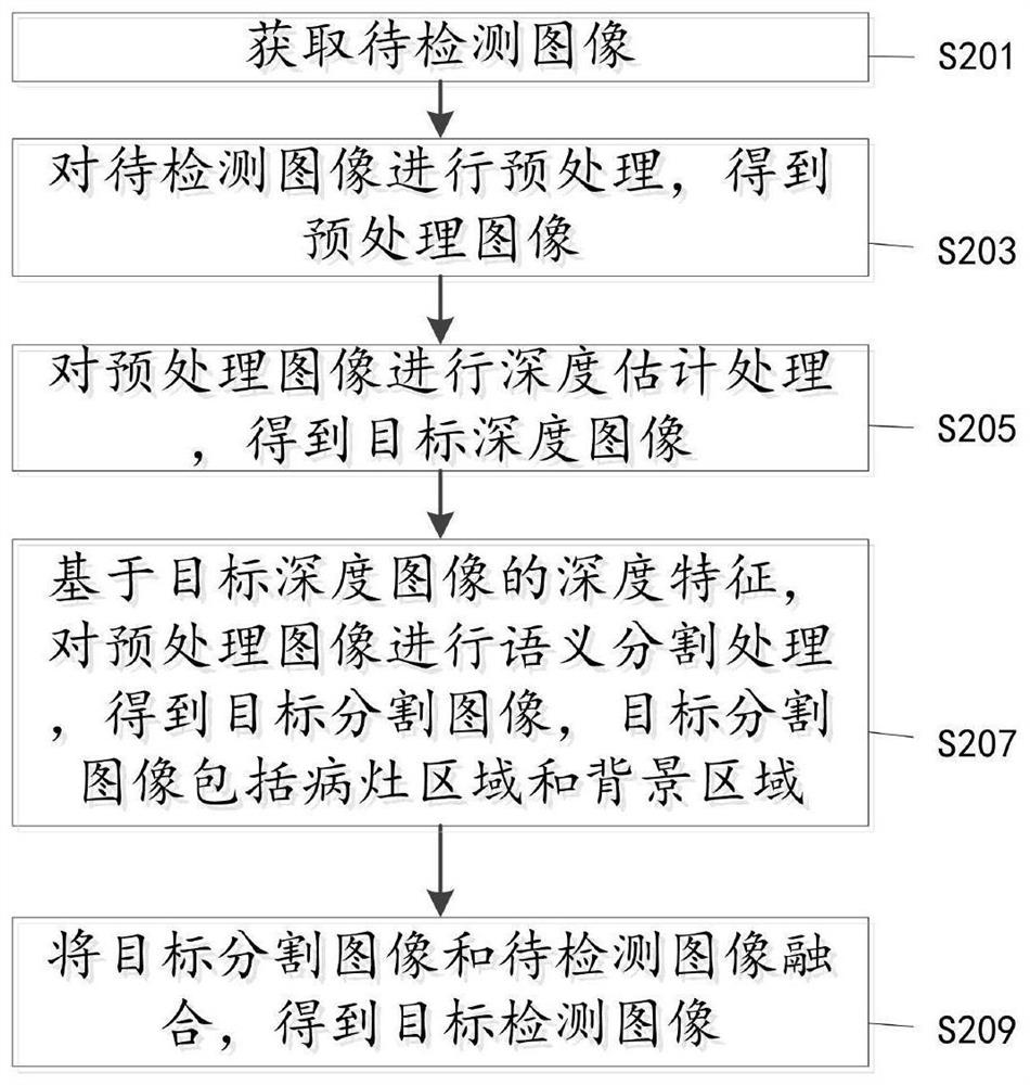
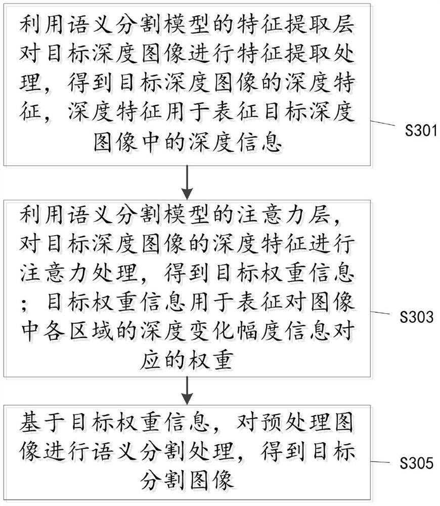
[0057]The following will clearly and completely describe the technical solutions in the embodiments of the application with reference to the drawings in the embodiments of the application. Apparently, the described embodiments are only some of the embodiments of the application, not all of them. Based on the embodiments in the present application, all other embodiments obtained by persons of ordinary skill in the art without making creative efforts belong to the protection scope of the present application.

[0058] It should be noted that the terms "first" and "second" in the description and claims of the present application and the above drawings are used to distinguish similar objects, but not necessarily used to describe a specific sequence or sequence. It is to be understood that the data so used are interchangeable under appropriate circumstances such that the embodiments of the application described herein can be practiced in sequences other than those illustrated or desc...


PUM


Abstract

The invention discloses an endoscopic image lesion area detection method, device and equipment and a storage medium. The method comprises the steps of obtaining a to-be-detected image; preprocessing the to-be-detected image to obtain a preprocessed image; performing depth estimation processing on the preprocessed image to obtain a target depth image; based on depth features of the target depth image, performing semantic segmentation processing on the preprocessed image to obtain a target segmented image, wherein the target segmented image comprises a focus area and a background area; and fusing the target segmented image and the to-be-detected image to obtain a target detection image. According to the application, the semantic segmentation model is guided to segment the pre-processed image through the obtained depth image, so that the detection rate and the detection accuracy of a tiny lesion area are improved, and the segmentation detection effect of the lesion area edge is improved, the false positive probability is greatly reduced, and a good auxiliary effect is achieved on clinical endoscopy.

Description

technical field [0001] The present application relates to the field of intelligent medical technology, and in particular to a method, device, equipment and storage medium for detecting lesion areas in endoscopic images. Background technique [0002] An endoscope is a detection instrument that integrates traditional optics, ergonomics, precision machinery, modern electronics, mathematics, and software; it has image sensors, optical lenses, light source lighting, and mechanical devices, etc. It can enter the stomach through the mouth Or enter the body through other natural channels. The endoscope can be used to see lesions that cannot be seen on X-rays, so it is very useful for doctors. For example, with the help of an endoscope, doctors can observe ulcers or tumors in the stomach, and formulate the best treatment plan accordingly; [0003] Endoscopy has always been a routine and important method for the examination of various diseases, and it has been widely used in the exa...

Claims


Application Information

Patent Timeline
IPC IPC(8): G06V10/26G06V10/44G06V10/774G06V10/80G06V10/82G06K9/62G06N3/04G06N3/08
CPCG06N3/08G06N3/045G06F18/214G06F18/253
Inventor 李凌陈宇桥徐强辜嘉李文超
Owner SUZHOU ZHONGKE HUAYING HEALTH TECH CO LTD
Why Eureka
Try Eureka