Image enhancement method and device for diabetic renal lesion classification

A kidney lesion and image enhancement technology, applied in the field of medical image processing, can solve problems such as low quality of CT images and limit the classification accuracy of image classification models, and achieve the effects of improving image quality, feature extraction and fusion, and improving efficiency

Pending Publication Date: 2022-02-11
成都市第二人民医院
View PDF0 Cites 3 Cited by
  • Summary
  • Abstract
  • Description
  • Claims
  • Application Information

AI Technical Summary

Problems solved by technology

However, due to the limitations of equipment performance and medical costs, the quality of the actu

Method used


Image

  • Image enhancement method and device for diabetic renal lesion classification
  • Image enhancement method and device for diabetic renal lesion classification
  • Image enhancement method and device for diabetic renal lesion classification

Examples


Example Embodiment

[0049] Example 1:

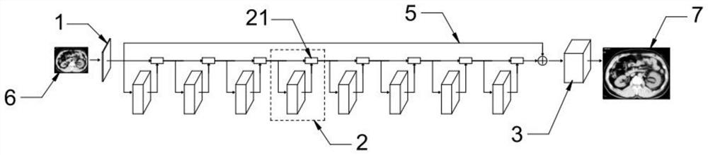
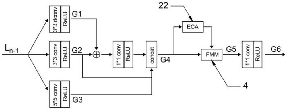
[0050] The super-resolution reconstruction network architecture and the structure of each module are as used in the structure of the present invention. Figure 1 - Figure 6 As shown, the MDC feature extracting unit 2 is set to eight, the convolution layer of the initial feature extracting unit 1 is 3 * 3, and the second image 6 of the super-division network is the three-channel image, and after the initial feature extracting unit 1, A first feature of the number of channels is obtained. In the MDC feature extraction unit 2, the number of G1, G2, and G3 feature drawing channels of the three branch extracted outputs are 48, G1 and G2 pass through the element, 1 * 1 volume and the RELU activation function, and G2, G3 splicing, giving a G4 feature of the channel number 144.

[0051] Inside the enhanced channel attention module 22, such as Figure 4 As shown, four branches are parallel relationships, and the four branches receive the G4 feature as input, each branch i...

Example Embodiment

[0056] Example 2:

[0057] As a comparative experiment, in the case where the relevant factors such as the control data set and the loss function are exactly the same, only the enhanced channel attention module 22, the modulation module 4 and the connection structure thereof and its connection structure thereof are provided in Example 1. Modify Figure 7 As shown, as the ratio I. In the ratio I, four branches spliced ​​after the feature map G4 modulated. The enhanced channel attention module 22, the modulation module 4 and the connection structure thereof and its connection structure thereof are modified as if the present invention will provide the super-resolution reconstruction network. Figure 8 As shown, as a contraction II. In contrast II, the third branch 25 in the enhanced channel focus module 22 is removed.

[0058] The reconstruction effect is measured after the same data set as in Example 1, and the comparative example II training is completed. The results show that in Exa...


PUM


Abstract

The invention discloses an image enhancement method for diabetic renal lesion classification and electronic equipment, the enhancement method comprises the steps of contrast enhancement, denoising, super-resolution reconstruction and the like, and the accuracy of an image classification network on diabetic renal lesion classification is improved by improving the quality of a kidney CT image. The super-resolution reconstruction network comprises a preliminary feature extraction unit, an MDC feature extraction unit and an up-sampling unit; the MDC feature extraction unit is arranged at the downstream end of the preliminary feature extraction unit; the plurality of MDC feature extraction units are sequentially connected end to end; and the up-sampling unit is arranged at the downstream end of the MDC feature extraction unit and is used for carrying out super-resolution reconstruction on the second feature map. According to the invention, the super-division network is used for repeatedly utilizing the features, the situation that low-layer feature information gradually disappears in continuous nonlinear operation is effectively avoided, and the model obtains good balance between the feature extraction effect and the complexity.

Description

technical field [0001] The invention belongs to the technical field of medical image processing, and in particular relates to an image enhancement method and equipment for classifying diabetic nephropathy. Background technique [0002] Diabetic nephropathy is a common complication of diabetes and one of the leading causes of death due to diabetes. Patients typically present with anorexia, weight loss, weakness, proteinuria, and hypertension. The traditional diagnostic method relies on doctors to judge the stage of diabetic nephropathy by visually observing the pathological images of the kidneys, and then formulate targeted treatment plans. However, the diagnosis of the disease depends heavily on the actual clinical experience of the doctor, and the workload is heavy, especially for some key hospitals, where there are many patients and the medical burden is heavy. [0003] Using image classification network to classify kidney CT images to assist in the diagnosis of diabetic...

Claims


Application Information

Patent Timeline
IPC IPC(8): G06T5/00G06T3/40G06N3/08G06N3/04G06K9/62
CPCG06T5/007G06T3/4053G06T5/002G06T3/4046G06T3/4038G06N3/08G06T2207/20081G06T2207/20084G06T2207/30084G06T2200/32G06N3/048G06N3/045G06F18/253Y02A90/10
Inventor 黄昶荃李永红李江玥孙玉彬
Owner 成都市第二人民医院
Why Eureka
Try Eureka