
Method for constructing in-situ primary bladder cancer animal model

A bladder and animal technology, applied in the field of tumor animal models, can solve the problems of short tumor formation time, inability to be used for tumor research, and long time-consuming

Pending Publication Date: 2022-02-22
WEST CHINA HOSPITAL SICHUAN UNIV
View PDF6 Cites 0 Cited by
  • Summary
  • Abstract
  • Description
  • Claims
  • Application Information

AI Technical Summary

Problems solved by technology

The genetic mouse model is a spontaneous model with a clear genetic background, but it is technically difficult and time-consuming; recently, Michael M.Shen's team constructed a mouse animal model of bladder cancer organoids derived from clinical patients, which has the advantages of Tumor organoids can retain the heterogeneity of the patient's tumor to a greater extent, and the tumor formation time is shorter. It can be used for research on tumor development and drug screening, but it cannot be used to study the process of tumor occurrence.
Looking at the above existing models, we still lack an animal model that can better simulate the occurrence and development of tumors in clinical patients

Method used


Image

  • Method for constructing in-situ primary bladder cancer animal model
  • Method for constructing in-situ primary bladder cancer animal model
  • Method for constructing in-situ primary bladder cancer animal model

Examples


Embodiment 1

[0068] Embodiment 1 Organoid culture of the present invention and method for expanding culture

[0069] Including obtaining fresh bladder tissue cells, trypsinization into single cells; in vitro 3D culture conditions to cultivate bladder tissue organoids.

[0070] method:

[0071] (1) Take 1-2 fresh mouse bladder tissues and cut them into pieces on ice;

[0072] (2) 8mL digestion solution (1.5mg / mL collagenase I and 1mg / mL collagenase IV+10uM Y27632) to resuspend the shredded tissue pieces (Y27632 can inhibit cell death and maintain cell activity during digestion);

[0073] (3) Digest the tissue resuspended in the digestive solution on a shaker at 37°C at a speed of 220rpm for 50min, pipetting once every 10min. Fully disperse the tissue cells;

[0074] (4) filter the digested tissue fluid with a 100 μm cell mesh;

[0075] (5) After filtration, centrifuge at room temperature, 1500rpm, and 5min to remove the supernatant;

[0076] (6) Add 3ml DMEM / F12 to resuspend, room temp...

Embodiment 2

[0090] Example 2 Gene Editing

[0091] The gene editing in this embodiment includes gene knockout and gene overexpression.

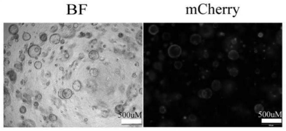
[0092] Gene knockout is: mouse bladder organoids are knocked out using CRISPR / cas9 technology. That is, the sgRNA vector and the Cas9 expression vector are packaged into a lentivirus, and then the lentivirus is transfected into an organoid, cultivated, and the gene knockout effect is verified by T7E1 enzyme. Among them, the sgRNA vector used in CRISPR / cas9 technology is pLenti-sgRNA-EFs-mcherry (with mCherry red fluorescent reporter gene).

[0093] Gene overexpression is: use the overexpression vector plasmid to perform gene overexpression in organoids, and the overexpression vector also contains a sequence for expressing luciferase. When cells are transferred into the vector plasmid, the cells can express luciferase. When the overexpression vector Bioluminescence is detected when the enzyme binds to its substrate (luciferin). The overexpression of ge...

experiment example 1

[0099] Experimental example 1 Construction of a muscle-invasive bladder cancer model

[0100] 1. Method

[0101] Using the methods in Examples 1 and 2, organoid culture, passage, and gene editing (combination A) were carried out sequentially, and the resulting gene-edited organoids were transplanted into the mouse bladder, and the size and location of the tumor were monitored by in vivo imaging technology, and transplanted for 30 Two days later, the mice were sacrificed, and bladder tissue was observed by direct observation, HE staining and immunohistochemical (IHC) staining.

[0102] 2. Results

[0103] 2.1 Organoid culture

[0104] Such as figure 2 As shown, individual bladder cells gradually grow into organoids over time.

[0105] 2.2 Identification of gene editing

[0106] The results of fluorescence detection after organoid gene editing are as follows: image 3 As shown in , visible organoids have been successfully transfected with viruses for gene editing.

[010...


PUM


Abstract

The invention discloses an in-situ primary bladder cancer tumor model preparation method, and belongs to the field of tumor animal models. According to the invention, normal bladder cells of mice are cultured into organoids with a specific culture medium, and then the organoids are subjected to gene editing and injected back to the bladder of the mice, so that the organoids are developed into tumors. Compared with a genetic engineering tumor animal model, the method disclosed by the invention is short in time consumption and high in tumor formation rate; compared with a transplanted tumor animal model, the model has an in-vivo microenvironment for tumor generation and development and a process from normal to tumor generation and development, and is closer to the most real state of bladder cancer generation and development.

Description

technical field [0001] The invention belongs to the field of tumor animal models. Background technique [0002] Bladder cancer (BC) is a common malignant tumor of the urinary system. According to the latest global statistics, in 2018, there were about 550,000 new cases of bladder cancer (3.0%) in the world, accounting for the twelfth place in tumor incidence, and about 200,000 cases (2.1%) of deaths, accounting for the fourteenth place in cancer mortality . The risk of bladder cancer in men is about 3-4 times higher than in women: bladder cancer is the sixth most common (4.5%) in men and ninth in women (2.8%). In my country, bladder cancer is the most common malignant tumor of the urinary system, and its incidence rate ranks first among male genitourinary system malignancies in China and is increasing year by year; among men, bladder cancer has risen to the sixth place in tumor incidence, and among women No. 11, it is bound to cause a huge burden on my country's medical an...

Claims


Application Information

Patent Timeline
IPC IPC(8): C12N5/071C12N15/90C12N15/867C12N15/85A01K67/027A61K49/00
CPCC12N5/0684C12N15/907C12N15/86C12N15/85A01K67/0271A61K49/0008C12N2501/415C12N2501/11C12N2533/90C12N2501/15C12N2500/38C12N2501/345C12N2740/15043C12N2800/107A01K2207/12A01K2227/105A01K2267/0331Y02A50/30
Inventor 陈崇王漫丽刘玉
Owner WEST CHINA HOSPITAL SICHUAN UNIV
Learn More