Method for constructing orthotopic primary endometrial cancer animal model

An endometrial cancer, animal model technology, applied in the fields of botanical equipment and methods, biochemical equipment and methods, animal cells, etc., can solve the problem of large differences in tumor pathophysiology, long preparation period, and can not meet the needs of model construction And other issues

Active Publication Date: 2022-02-22
WEST CHINA HOSPITAL SICHUAN UNIV
View PDF1 Cites 3 Cited by
  • Summary
  • Abstract
  • Description
  • Claims
  • Application Information

AI Technical Summary

Problems solved by technology

[0004] The genetically engineered animal model has a good tumor microenvironment and good reproducibility, and the immune system is not defective, but it needs to produce transgenic animals, which is costly and takes a long time to prepare
The cell line xenograft tumor model only needs to implant the human tumor cell line into the model animal, which is easy to prepare and has high reproducibility, but it needs to use immunodeficient mice, and the obtained tumor is not the primary tumor in situ, which is different from the actual development of the tumor. The situation is quite different from the pathophysiological situation
The PDX model is to inoculate the patient's tumor tissue into the model animal body. It is easy to prepare and the genotype is close to the actual tumor. However, the non-situ tumor cannot provide the in situ microenvironment of the endometrium tissue, which may lead to the occurrence of human tumors in the experimental process. The relevant biological characteristics in the middle are lost, and the situation in the human body cannot be simulated. Moreover, clinical tumor specimens are very precious at present. Some special clinical specimens such as puncture specimens and other small specimens have less tissue cells for experimental research. The PDX model of endometrial cancer The construction success rate is also relatively low, which cannot meet the model construction requirements

Method used


Image

  • Method for constructing orthotopic primary endometrial cancer animal model
  • Method for constructing orthotopic primary endometrial cancer animal model
  • Method for constructing orthotopic primary endometrial cancer animal model

Examples


Embodiment 1

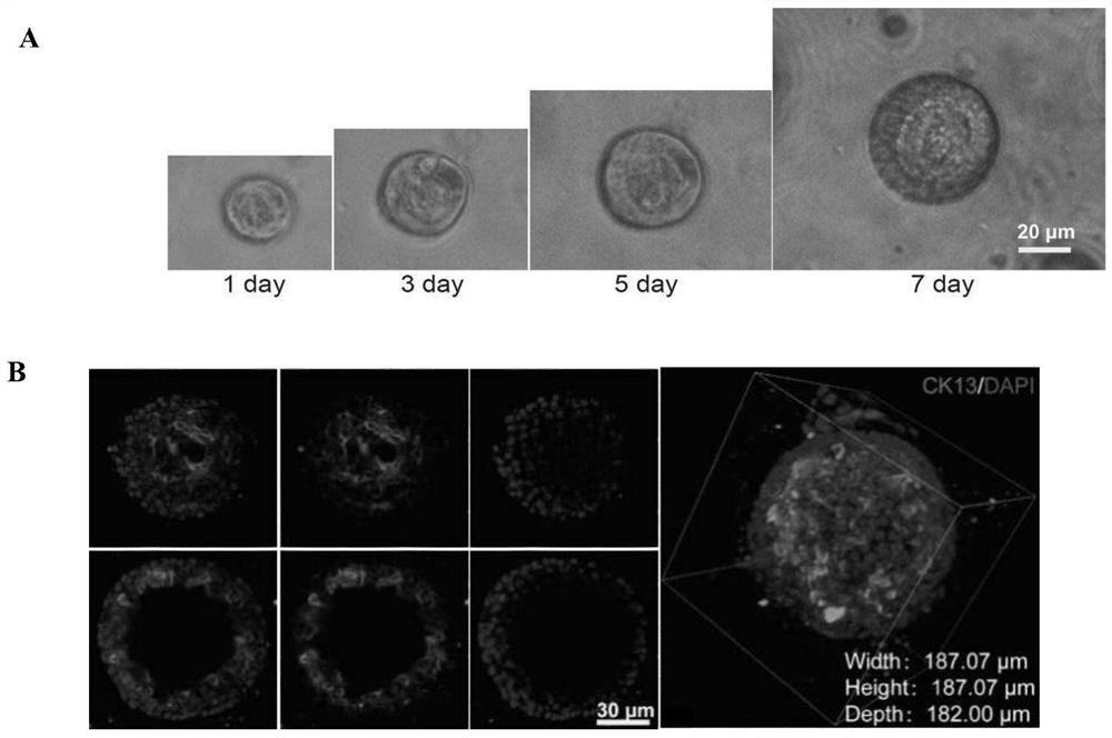
[0076] Embodiment 1 Organoid culture method of the present invention

[0077] It includes the following steps:

[0078] (1) Take fresh mouse endometrium and cut it on ice;

[0079] (2) Collagenase (2mg / mL Collagenase I and 1mg / mL Collagenase IV) to resuspend the chopped tissue blocks, and use the gentleMACS automatic tissue processor to run Mouse Tumor program 1 in the C tube; chopped tissue blocks The dosage is 1-2 grams, and the dosage of collagenase is 10 mL;

[0080] (3) The tissue block treated with collagenase was shaken at 37°C and the speed was 220rpm for 30min digestion. Make the tissue cells fully dispersed;

[0081] (4) Transfer the digested solution to the automatic tissue processor gentleMACS. On the gentleMACS, run the Mouse Tumor program 2;

[0082] (5) Filtering the liquid containing endometrial cells processed in step 4 with a 100 μm cell mesh;

[0083] (6) after filtration, at room temperature, 1500rpm, 5min centrifugation to remove supernatant;

[008...

Embodiment 2

[0093] Example 2 The modeling method of the present invention

[0094] 1. Modeling method

[0095] 1.1 Pre-culture

[0096] Same as Example 1.

[0097] 1.2 Expanding the culture

[0098] (1) Take the organoids that have been cultured for about 7 days, resuspend the digested organoids with TrypLE, transfer them to a 15mL centrifuge tube, add 3ml TrypLE to one well of a 48-well plate, and pipet 10 to 20 times until the matrigel is completely broken. , digested in a water bath at 37°C for 5 min;

[0099] (2) Take out from the water bath, pipet again for 20 to 30 times, digest at 37°C for 5 min, and then perform the third pipetting (20 to 30 times). Digestion into single cells when viewing organoids under a microscope. If a single cell is not formed, the water bath and pipetting can be repeated once until a single cell is formed.

[0100] (3) 1500rpm, centrifuge at room temperature for 5min, remove the supernatant;

[0101] (4) After counting the cells, add 30 μL of Martrig...

experiment example 1

[0135] Experimental example 1 Construction of a kind of endometrial adenocarcinoma model

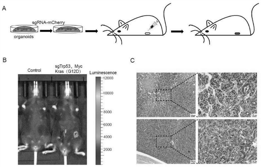
[0136] The endometrial cancer model was constructed using the method in Example 2, wherein gene combination 1 in Table 2 was selected for the gene editing part. That is, endometrial cancer organoids with knockout of Trp53 gene and overexpression of Kras(G12D) and Myc were cultured, orthotopically transplanted into the uterus of mice to construct an animal model of endometrial cancer. 135 days after transplantation, all three mice developed tumors.

[0137] The schematic diagram of the construction process is as follows figure 2 As shown in A.

[0138] figure 2 B shows the live imaging results of tumors in mice 95 days after transplantation. It can be seen that tumors formed in the uterus of mice transformed with gene-edited organoids.

[0139] figure 2 C shows that the tumor exhibits features of poorly differentiated adenocarcinoma.


PUM


Abstract

The invention discloses an in-situ primary endometrial cancer tumor model preparation method, and belongs to the field of tumor animal models. The method comprises the following steps: culturing mouse endometrial cells into organoids by using a specific culture medium, performing gene editing on the organoids, and injecting the organoids back to the mouse endometrium so as to enable the organoids to develop into tumors. The tumor model constructed by the invention has the characteristics of primary, in-situ and definite genotype, and compared with a genetic engineering tumor animal model, the time consumption is short, and the tumor formation rate is high; compared with a transplanted tumor animal model, the model has an in-vivo microenvironment for tumor generation and development, is closer to the most real state of endometrial cancer, and has a good application prospect.

Description

technical field [0001] The invention belongs to the field of tumor animal models. Background technique [0002] Endometrial cancer is a group of epithelial malignant tumors that occur in the endometrium, and it is more common in perimenopausal and postmenopausal women. Endometrial cancer is one of the most common female reproductive system tumors, with nearly 200,000 new cases each year, and is the third most common gynecological malignancy that causes death (after ovarian cancer and cervical cancer). Its incidence is closely related to lifestyle, and its incidence varies in different regions. In North America and Europe, its incidence is second only to breast cancer, lung cancer, and colorectal tumors, ranking first among female reproductive system cancers. In my country, with the development of society and the improvement of economic conditions, the incidence of endometrial cancer is also increasing year by year, ranking second only to cervical cancer, ranking second amon...

Claims


Application Information

Patent Timeline
Patent Type & Authority Applications(China)
IPC IPC(8): C12N5/071C12N15/867C12N15/12C12N15/65A01K67/027A61K49/00
CPCC12N5/0682C12N15/86C12N15/65C07K14/82C07K14/4703A01K67/0271A61K49/0008C12N2509/00C12N2501/11C12N2501/119C12N2501/415C12N2501/998C12N2501/727C12N2501/999C12N2501/345C12N2501/18C12N2500/32C12N2501/15C12N2500/38C12N2501/392C12N2740/15043C12N2800/107A01K2207/12A01K2227/105A01K2267/03A01K2267/0393
Inventor 陈崇陈婧瑶刘玉
Owner WEST CHINA HOSPITAL SICHUAN UNIV
Why Eureka
Try Eureka