Enhanced CT (Computed Tomography) imageomics feature processing method for upper abdomen of liver cirrhosis patient and application thereof

A technology of CT imaging and radiomics, applied in the fields of medicine, nuclear medicine and radiomics, can solve the problems of doctors' reading and interpretation, high price, and low accuracy

Pending Publication Date: 2022-03-01
BEIJING YOUAN HOSPITAL CAPITAL MEDICAL UNIV +1
View PDF0 Cites 2 Cited by
  • Summary
  • Abstract
  • Description
  • Claims
  • Application Information

AI Technical Summary

Problems solved by technology

However, HVPG measurement is an invasive angiographic intervention that requires specialized equipment and is relatively expensive; at the same time, it is easily affected by some factors: such as body position, breathing, anesthetic drugs and intrahepatic shunt, etc.
Some scholars have proposed several diagnostic and predictive models based on serum markers, such as FIB-4 (Fibrosis Index Based on Four Factors, liver fibrosis index), Lok score, APRI (Aspartate aminotransferase to platelet count ratio index, aspartate aminotransferase to platelet count ratio index, aspartate aminotransferase and Platelet count ratio index), AAR (Aspartate aminotransferase to alanine aminotransferaseratio, ratio of aspartate aminotransferase to alanine aminotransferase), VITRO score (Von Willebrand factor antigen / platelet ratio, ratio of von Willebrand factor antigen level to platelet count), etc., but These models are not very accurate
[0005] In CT imaging examination, in addition to the size, shape, edge shape, enhanced structure and other characteristics of organs or lesions that can be observed by the naked eye, a large amount of image data will be generated at the same time, and these data cannot be completely read and interpreted by doctors

Method used


Image

  • Enhanced CT (Computed Tomography) imageomics feature processing method for upper abdomen of liver cirrhosis patient and application thereof
  • Enhanced CT (Computed Tomography) imageomics feature processing method for upper abdomen of liver cirrhosis patient and application thereof
  • Enhanced CT (Computed Tomography) imageomics feature processing method for upper abdomen of liver cirrhosis patient and application thereof

Examples


Embodiment 1

[0096] Example 1 Basic information of patients with liver cirrhosis included in the study

[0097] 1.1 Research object

[0098] The clinical data, laboratory data, gastroscopy and enhanced CT images of the upper abdomen were collected retrospectively from January 2016 to December 2017 of inpatients with liver cirrhosis in Beijing You'an Hospital affiliated to Capital Medical University. The diagnosis of liver cirrhosis is mainly based on medical history, pathological tissue biopsy, typical clinical symptoms, laboratory tests and imaging findings. The diagnostic criteria for liver cirrhosis in this study were in line with the guidelines for the diagnosis and treatment of liver cirrhosis (2019 edition).

[0099] Inclusion criteria include: (1) aged 18-85 years; (2) gender is not limited; (3) meet the diagnostic criteria for liver cirrhosis, the etiology is not limited; (4) completed gastroscopy and upper abdominal enhanced CT examination, and both The time difference shall n...

Embodiment 2

[0109] Example 2 A method for image-omics feature processing of enhanced abdominal CT in patients with liver cirrhosis

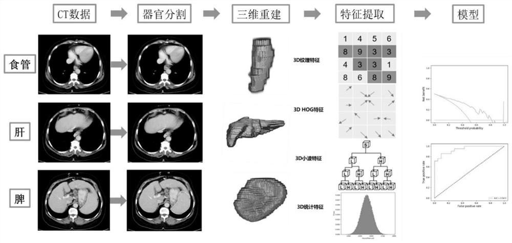
[0110] The flow process of the method described in this embodiment is as follows figure 1 As shown, based on the research object described in embodiment 1, the specific establishment process is:

[0111] S1. Collect enhanced CT images of the upper abdomen of patients with liver cirrhosis, and upload the CT images in the portal venous phase with the most obvious enhancement of the liver, spleen and esophageal veins to the precision medicine open platform (developed by Beijing Yinji Information Technology (Beijing) Co., Ltd.) in DICOM format.

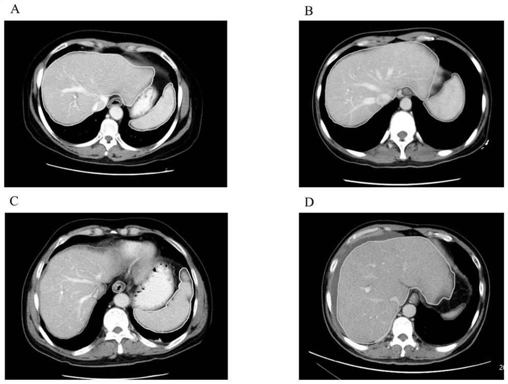
[0112] S2. First, the doctor marks the liver, spleen and lower esophagus in the enhanced CT image obtained in step S1 layer by layer. Since the most serious part of varicose veins mostly occurs within 5 cm above the cardia, the marking range of the esophagus is the lower esophagus, from the cardia to its upper 5 cm. ...


PUM


Abstract

The invention provides a processing method for enhancing CT (Computed Tomography) image omics characteristics of the upper abdomen of a liver cirrhosis patient and application of the processing method in judging esophageal varicosity of the liver cirrhosis patient. The processing method comprises the following steps: S1, collecting an enhanced CT image of the upper abdomen of a liver cirrhosis patient; s2, marking the lower section areas of the liver, the spleen and the esophagus in the enhanced CT image layer by layer; s3, performing three-dimensional reconstruction on the marked areas respectively; s4, image omics features are extracted from each three-dimensional area and then fused, and original image omics features of the patient are obtained; s5, performing dimensionality reduction processing on the original radiomics characteristics by applying a principal component analysis method to obtain dimensionality-reduced radiomics characteristics; and S6, constructing a patient esophageal varicosity radiomics classification model by using a classification algorithm.

Description

technical field [0001] The invention relates to the fields of medicine, nuclear medicine and radiomics, in particular to a method for processing radiomics features of enhanced abdominal CT in patients with liver cirrhosis and its application in judging esophageal varices in patients with liver cirrhosis. Background technique [0002] Cirrhosis is the terminal stage of progression of various chronic liver diseases and is the 11th most common cause of death worldwide. Variceal bleeding is a serious and fatal complication of liver cirrhosis. Esophageal varices have a higher mortality rate than gastric varices. About 1 / 3 of patients with esophageal varices will experience bleeding from esophageal varices, and the mortality rate after acute hemorrhage is as high as 15%-25% within 6 weeks, and as high as 40% within 1 year after hemorrhage. Therefore, early diagnosis of varices, especially esophageal varices, prediction of bleeding risk, and selection of appropriate treatment are...

Claims


Application Information

Patent Timeline
Patent Type & Authority Applications(China)
IPC IPC(8): G16H30/20G16H30/40G06T11/00G06V10/50G06V10/77G06V10/764G06V10/80G06K9/62
CPCG16H30/20G16H30/40G06T11/003G06T2211/416G06F18/2135G06F18/2411G06F18/253
Inventor 范春蕾刘瑾
Owner BEIJING YOUAN HOSPITAL CAPITAL MEDICAL UNIV
Why Eureka
Try Eureka