Deep learning-based CT image endangered organ segmentation system

A CT image and deep learning technology, applied in the field of medical image processing, can solve problems such as reducing the level of network nonlinearity, pixel misclassification, and poor segmentation effect, so as to improve segmentation accuracy, strengthen learning ability, and enhance nonlinearity. Effect

Active Publication Date: 2022-03-22
SOUTH CHINA UNIV OF TECH
View PDF5 Cites 6 Cited by
  • Summary
  • Abstract
  • Description
  • Claims
  • Application Information

AI Technical Summary

Problems solved by technology

[0004] 1. Lack of extraction and utilization of multi-scale information, the segmentation effect is not good for segmentation objects with complex structures or multiple segmentation objects with different structural sizes, and the segmentation accuracy of organs with small sizes is low
[0005] 2. In the process of restoring the size of the feature map by the segmentation system, there is a lack of utilization of global information, and the global information collecte

Method used


Image

  • Deep learning-based CT image endangered organ segmentation system
  • Deep learning-based CT image endangered organ segmentation system
  • Deep learning-based CT image endangered organ segmentation system

Examples


Embodiment Construction

[0050] The present invention will be further described in detail below in conjunction with the embodiments and the accompanying drawings, but the embodiments of the present invention are not limited thereto.

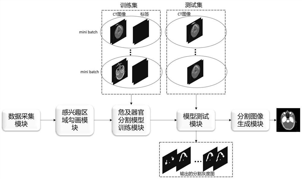
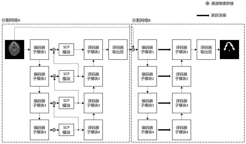
[0051] Such as figure 1 As shown, the deep learning-based CT image organ-at-risk segmentation system provided in this embodiment includes: a data acquisition module, a region-of-interest delineation module, an organ-at-risk segmentation model training module, a model testing module, and a segmented image generation module.

[0052] The data collection module is used to collect a CT image data set, and the CT image data set includes CT images containing multiple organs at risk of nasopharyngeal carcinoma. In this embodiment, the target part of the sample selection is concentrated on the head and neck due to pathological reasons; the CT image data set is randomly divided to form a training set and a test set, with a ratio of 4:1.

[0053] The region of interest delineatio...


PUM


Abstract

The invention discloses a CT image endangered organ segmentation system based on deep learning. The CT image endangered organ segmentation system comprises a data acquisition module, a region-of-interest sketching module, an endangered organ segmentation model training module, a model test module and a segmented image generation module. A pyramid-type deep learning network integrating global information flow and an SCP module located on jump connection and used for extracting and fusing multi-scale information are provided, the weight for segmenting useful features is increased through utilization of the multi-scale global information flow and an attention mechanism, the nonlinearity of the structure is enhanced, the performance of a segmentation model is remarkably improved, and the segmentation efficiency is improved. Meanwhile, a cascade network structure based on an automatic context method is designed, after the positioning result of the organ region to be segmented is combined with the original CT image input by using the automatic context method, refined segmentation is carried out, and the segmentation accuracy of the whole system is remarkably improved.

Description

technical field [0001] The present invention relates to the technical field of medical image processing, in particular to a CT image organ-at-risk segmentation system based on deep learning. Background technique [0002] Nasopharyngeal carcinoma is one of the malignant tumors with high incidence in China, and its incidence rate is the first among malignant tumors of the ear, nose and throat. When patients are treated with radiation therapy for this type of tumor, if the radiation target area cannot be strictly controlled, it may endanger the Many normal organs and tissues are obtained, which will have adverse effects on the health of patients. CT images are the standard image resources for delineating target areas and organs at risk in radiotherapy. The corresponding target areas and organs are manually delineated by experienced physicians to clearly show their respective areas, so that the radiotherapy area is strictly controlled In the target area, normal organs will not ...

Claims


Application Information

Patent Timeline
IPC IPC(8): G06V10/26G06V10/25G06V10/764G06V10/82G06K9/62G06N3/04G06N3/08
CPCG06N3/08G06T7/11G06T7/0012G06T2207/20081G06T2207/20084G06T2207/30096G06T2207/10081G06N3/045G06F18/2415G06F18/2431Y02A90/10
Inventor 郭礼华黄泽曦
Owner SOUTH CHINA UNIV OF TECH
Why Eureka
Try Eureka