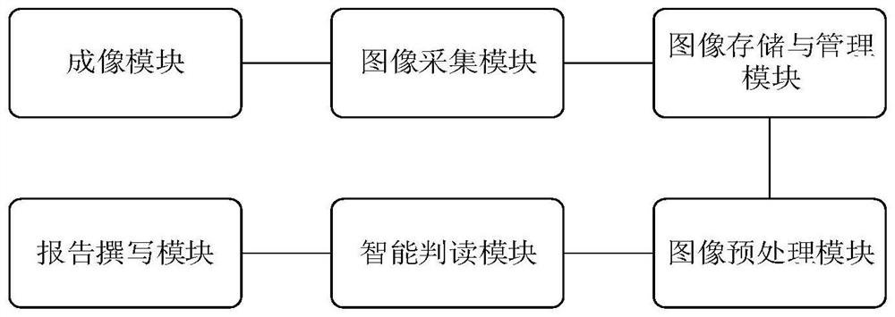
Automatic interpretation system for cell pathology smear
A cytopathology and smear technology, applied in microscopy, optics, biological neural network models, etc., can solve the problem of inability to distinguish benign and malignant specimens, and achieve the effect of reducing workload, improving accuracy, and improving accuracy
- Summary
- Abstract
- Description
- Claims
- Application Information
AI Technical Summary
Problems solved by technology
Method used
Image
Examples
Embodiment 1
[0096] In this embodiment, the digital microscope has a maximum 100 times optical zoom function, and the host computer is a computer with an X86 architecture.
[0097] 1. Obtain the training set
[0098] 1-1. Collect healthy cells (negative, atypical) and lung cancer cells and place them on glass slides, stain them with diffquick technology, and make cytopathological smears. The types of lesions are further divided into adenocarcinoma, squamous cell carcinoma, small cell carcinoma and large cell carcinoma. Cellular neuroendocrine carcinoma. refer to Figure 4 , Arrows indicate adenocarcinoma cells (blue).
[0099] 1-2. Place the cytopathological smear on the motorized stage, and the imaging module and image acquisition module work:
[0100] First, control the motorized stage to move until its center coincides with the central axis of the objective lens of the digital microscope, then complete the autofocus through the focus control module, and then fix the focal length; con...
PUM
 Abstract
Description
Claims
Application Information
 - R&D
- Intellectual Property
- Life Sciences
- Materials
- Tech Scout
- Unparalleled Data Quality
- Higher Quality Content
- 60% Fewer Hallucinations
Browse by: Latest US Patents, China's latest patents, Technical Efficacy Thesaurus, Application Domain, Technology Topic, Popular Technical Reports.
© 2025 PatSnap. All rights reserved.Legal|Privacy policy|Modern Slavery Act Transparency Statement|Sitemap|About US| Contact US: help@patsnap.com


