Eureka AIR delivers breakthrough ideas for toughest innovation challenges, trusted by R&D personnel around the world.

Fundus lesion recognition method and device, electronic equipment and readable storage medium

A recognition method, fundus image technology, applied in neural learning methods, character and pattern recognition, image analysis, etc., to achieve the effect of low-cost, low-model training and application

Pending Publication Date: 2022-05-17
ALIBABA (CHINA) CO LTD
View PDF0 Cites 4 Cited by
  • Summary
  • Abstract
  • Description
  • Claims
  • Application Information

AI Technical Summary

Problems solved by technology

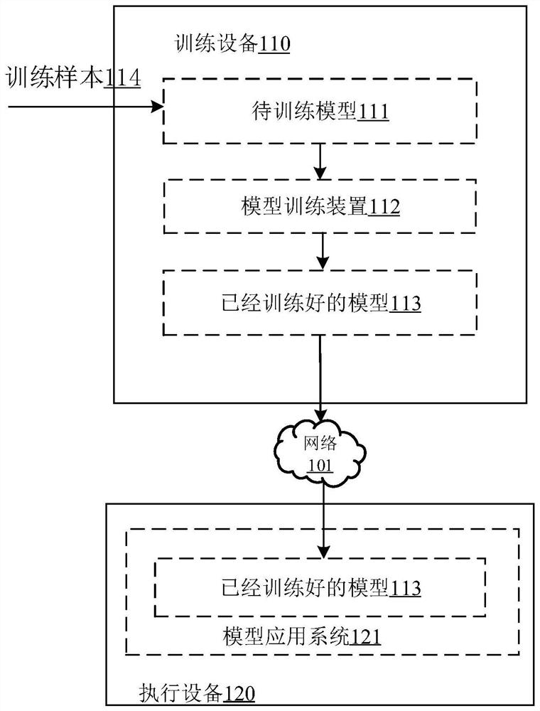
[0004] However, the current detection and diagnosis of fundus lesions based on OCT fundus images combined with deep learning models requires careful labeling of lesion locations on OCT fundus images (target boxes or densely labeled at the pixel level), and most clinical OCT fundus images only have Labeling of the patient's diagnostic level (that is, only the fundus lesions of the patient are marked on the OCT fundus image of a patient, and the target frame will not be marked in detail on the map)

Method used


Image

  • Fundus lesion recognition method and device, electronic equipment and readable storage medium
  • Fundus lesion recognition method and device, electronic equipment and readable storage medium
  • Fundus lesion recognition method and device, electronic equipment and readable storage medium

Examples


Embodiment Construction

[0038] The present disclosure is described below based on examples, but the present disclosure is not limited only to these examples. In the following detailed description of the disclosure, some specific details are set forth in detail. The present disclosure can be fully understood by those skilled in the art without the description of these detailed parts. In order to avoid obscuring the essence of the present disclosure, well-known methods, procedures, and procedures are not described in detail. Additionally, the drawings are not necessarily drawn to scale.

[0039] Before the specific introduction of each embodiment of the present disclosure, some of the knowledge used therein will be explained as follows.

[0040] The layered tissue structure of retinal tissue is: inner limiting membrane, nerve fiber layer, ganglion cell layer, inner sub-like layer, inner granular layer, outer sub-like layer, outer granular layer, inner segment, outer segment and retina Pigment epithe...


PUM


Abstract

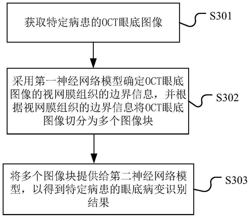
The invention provides a fundus lesion recognition method and device. The method comprises the following steps: acquiring an OCT fundus image of a specific patient; determining boundary information of a retina tissue of the OCT eye fundus image by adopting a first neural network model, and segmenting the OCT eye fundus image into a plurality of image blocks according to the boundary information of the retina tissue; the plurality of image blocks are provided for a second neural network model to obtain the fundus lesion type of the specific patient, and the second neural network model calculates the feature vector of each image block and the attention weight of each image block for the set fundus lesion type; and performing weighted calculation to obtain a fused feature vector so as to obtain an identification result aiming at a set fundus lesion type. Wherein the second neural network model reinforces important information in the image block group in combination with the spatial domain significance and the scanning position significance, and filters unimportant information, so that low-cost model training and application are realized.

Description

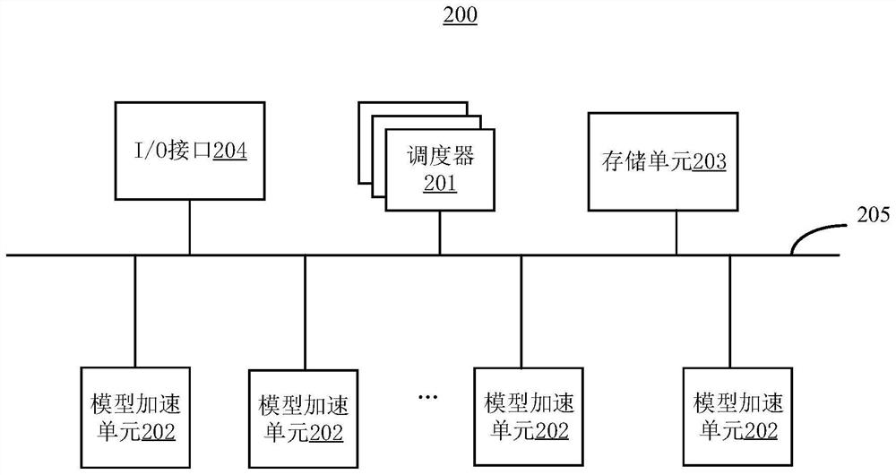
technical field [0001] The present disclosure relates to the technical field of combining artificial intelligence and medical image processing, and in particular to a fundus lesion identification method, device, electronic equipment and readable storage medium. Background technique [0002] Optical coherence tomography (OCT) imaging technology can scan the layered tissue structure of the retina in detail to obtain OCT fundus images that reflect the shape and thickness of each layer of the retinal layer. On the basis of OCT fundus images, it can assist doctors in the diagnosis of fundus lesions by intelligently processing the images, which can not only improve the efficiency of doctors' diagnosis of diseases to a certain extent, but also effectively improve the accuracy of disease diagnosis. [0003] In recent years, with the rise of deep learning, researchers have begun to apply neural network models to the medical field, and deep learning models have also been widely used i...

Claims


Application Information

Patent Timeline
IPC IPC(8): G06V10/764G06V10/80G06V10/82G06K9/62G06N3/04G06N3/08G06T7/00
CPCG06N3/08G06T7/0012G06T2207/10101G06T2207/20081G06T2207/20084G06T2207/30041G06T2207/30096G06N3/045G06F18/2415G06F18/253
Inventor 崔波陈漠沙邵天越王一婷
Owner ALIBABA (CHINA) CO LTD
Why Eureka
Eureka Blog
Learn More