Method for positioning pulmonary nodules

A positioning method and technology for small nodules, which can be used in radiological diagnosis instruments, medical science, surgery, etc., can solve the problems of puncture complications, patient pain, and high costs, so as to reduce complications, reduce positioning steps, and reduce The effect of positioning costs

Pending Publication Date: 2022-05-31
闾夏轶 +1
View PDF1 Cites 1 Cited by
  • Summary
  • Abstract
  • Description
  • Claims
  • Application Information

AI Technical Summary

Problems solved by technology

However, this positioning method has the following defects: (1) The selection of the puncture path is blocked by the bony structure of the thorax, and it is difficult to ensure that the puncture needle can be completely perpendicular to the surface of the pleura, resulting in the deviation of the resection site in the book; (2) After the Hook-Wire is implanted in the body, there is a risk of falling off and displacement due to the movement of the lung tissue; (3) The Hook-Wire is an invasive examination and requires repeated CT scans to confirm the position, and there are risks of radiation exposure and puncture complications (4) The puncture needle is left in the body, causing the patient's pain and discomfort
However, this method must rely on computer modeling and 3D printing of a strip-shaped positioning device, and the positioning device needs to be embedded in the chest cavity of the recipient during the operation, which is cumbersome and costly, and increases the risk of surgery. risk of infection
In addition, the applicable conditions are limited, for example, for pulmonary nodules located near the top and bottom of the lung and close to the heart, this method is difficult to apply due to the positional relationship

Method used


Image

  • Method for positioning pulmonary nodules
  • Method for positioning pulmonary nodules
  • Method for positioning pulmonary nodules

Examples


Embodiment 1

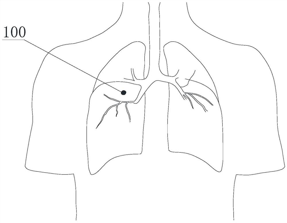
[0047] like figure 1 As shown, a pulmonary nodule 100 located deep in the middle lobe of the right lung (the vertical distance under the pleura is more than 5 mm) is taken as an example for illustration. The location of the small nodule is deep, and it is difficult to locate by vision, finger palpation, and strong instrument sliding during the operation, but it can be located by the positioning method provided by the present invention. Specific steps are as follows:

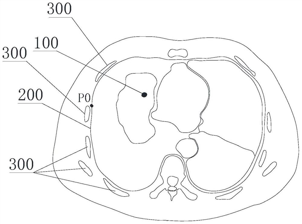
[0048] S0: Determine the location of the small pulmonary nodule in the lung: First, determine the specific location of the small pulmonary nodule in the lung according to the preoperative CT image, such as: at the height of the fifth rib on the transverse plane, and at the level of the 5th rib on the sagittal plane 10cm from the midline and 15cm from the anterior chest wall;

[0049] S1: Establishment of a 3D model of the thorax: According to the preoperative CT image, 3D modeling is performed by a computer, an...

Embodiment 2

[0075] The difference between this embodiment and Embodiment 1 is:

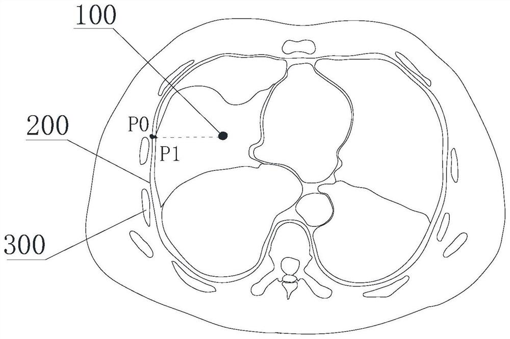
[0076] On the basis of Example 1, the virtual puncture location point P1 on the lung surface confirmed by S3 is checked and calibrated, and the operator determines the real chest wall projection point P2 of the small pulmonary nodule under direct vision of the thoracoscope according to the traditional location method. , compare with the virtual puncture location point P1 on the lung surface determined in step S3, check the validity of the virtual puncture location point P1 on the lung surface, and calibrate the model.

[0077] Traditional localization methods include locating small pulmonary nodules through the operator's vision and touch.

[0078] The above test and calibration are suitable for the case where the nodule is close to the lung surface. When the nodule is far away from the lung surface or it is difficult to locate according to the traditional positioning method due to anatomical reasons, the tes...


PUM


Abstract

The invention discloses a pulmonary nodule positioning method. The method comprises the following steps: S0, determining the position of a pulmonary nodule in a lung; s1, establishing a thoracic three-dimensional model; s2, marking a virtual puncture positioning point; s3, marking the position of the pulmonary nodule on the surface of the lung. According to the positioning method of the pulmonary nodule, accurate positioning and real-time registration are performed on the CT three-dimensional reconstruction image and the thoracoscope image by virtue of a computer-aided diagnosis technology and a computer deep learning model based on artificial intelligence, so that accurate positioning of the pulmonary nodule is realized. Meanwhile, interventional or radiative positioning steps harmful to the human body of the patient are reduced, complications after positioning can be reduced, and the positioning cost is reduced.

Description

technical field [0001] The invention relates to a method for locating small pulmonary nodules, which is used for assisting in locating small pulmonary nodules during an operation. Background technique [0002] Lung cancer is the malignant tumor with the highest morbidity and mortality in the world, which seriously threatens human life and health. China is a country with a high incidence of lung cancer, with both morbidity and mortality ranking first in the world. In recent years, although the diagnosis and treatment technology of lung cancer has made great progress, the overall survival rate of lung cancer patients is still not optimistic. The main reason for the poor prognosis of lung cancer patients is that most patients have already reached an advanced stage when their symptoms are discovered. Therefore, early diagnosis and early treatment of lung cancer are the core links in the prevention and treatment of lung cancer. Chest low-dose helical CT screening can detect sma...

Claims


Application Information

Patent Timeline
Patent Type & Authority Applications(China)
IPC IPC(8): A61B34/10A61B6/03A61B6/00
CPCA61B34/10A61B6/032A61B6/5247A61B2034/105A61B2034/107A61B2034/108
Inventor 闾夏轶
Owner 闾夏轶
Why Eureka
Try Eureka