Sample component analysis method, apparatus and device, and storage medium

A component analysis and sample technology, applied in the field of image processing, can solve problems such as incomplete image information, and achieve the effect of ensuring accuracy, reducing false recognition rate and missed recognition rate

Pending Publication Date: 2022-07-29
COBIO SMARTER HEALTHCARE TECH
View PDF0 Cites 1 Cited by
  • Summary
  • Abstract
  • Description
  • Claims
  • Application Information

AI Technical Summary

Problems solved by technology

[0004] The present invention provides a sample composition analysis method, device, equipment and storage medium to solve the problem of incomplete image information of existing target images and improve the accuracy of sample analysis results

Method used


Image

  • Sample component analysis method, apparatus and device, and storage medium
  • Sample component analysis method, apparatus and device, and storage medium
  • Sample component analysis method, apparatus and device, and storage medium

Examples


Embodiment 1

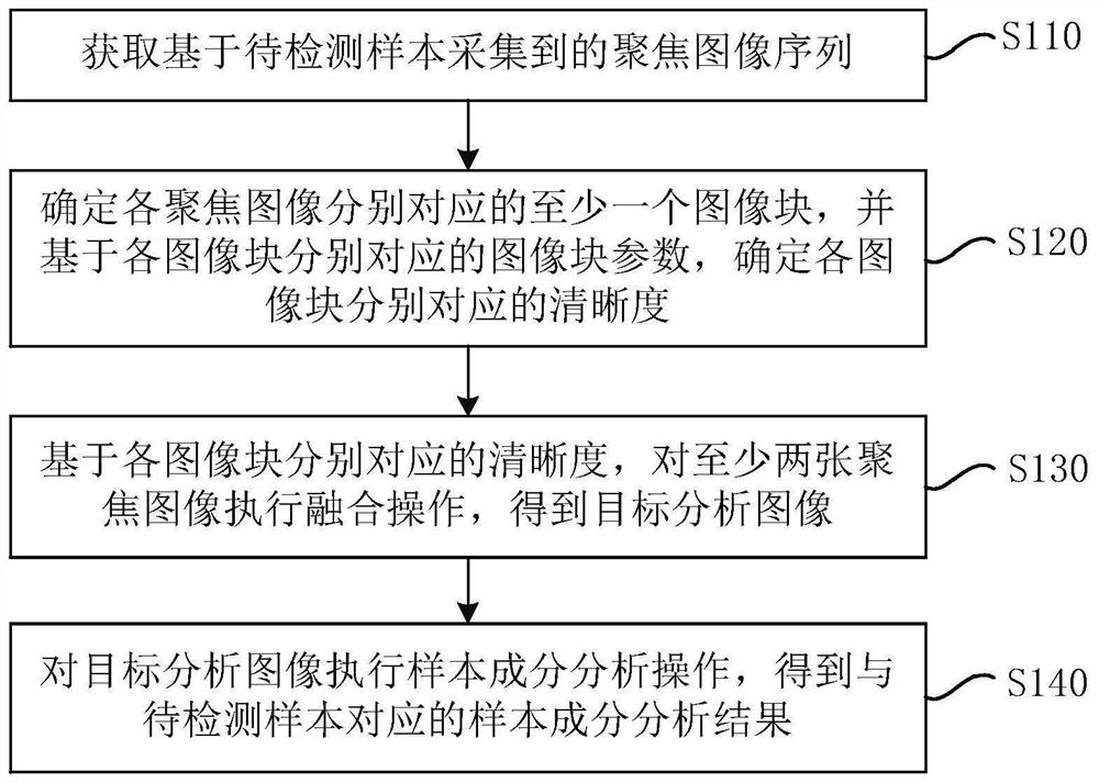
[0035] figure 1 This is a flow chart of a method for analyzing sample components according to Embodiment 1 of the present invention. This embodiment can be applied to the case of performing sample component analysis on a body fluid sample. The method can be executed by a sample component analyzing device. The sample component analysis The device can be implemented in the form of hardware and / or software, and the sample component analysis device can be configured in a terminal device or a sample detection device. like figure 1 As shown, the method includes:

[0036] S110. Acquire a focused image sequence collected based on the sample to be detected.

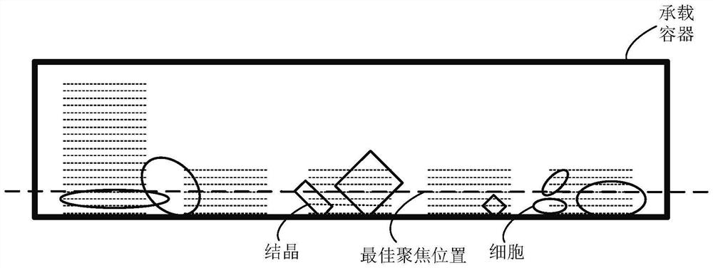
[0037] Exemplarily, the sample to be detected may be a body fluid, and the body fluid refers to a liquid substance in the body. Specifically, the body fluid includes but is not limited to blood, tissue fluid, lymph fluid, urine, pleural and ascites fluid, and cerebrospinal fluid. The liquid composition in the sample to be detec...

Embodiment 2

[0066] Figure 5 This is a flowchart of a sample component analysis method provided according to the second embodiment of the present invention. This embodiment further refines the "determining target analysis image based on at least two fused image blocks" in the above embodiment. like Figure 5 As shown, the method includes:

[0067] S210. Acquire a focused image sequence collected based on the sample to be detected.

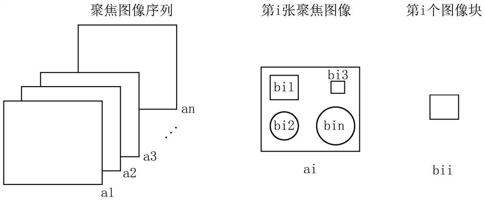
[0068] S220: Determine at least one image block corresponding to each focused image, and determine the resolution corresponding to each image block based on the image block parameters corresponding to each image block.

[0069] In an embodiment, optionally, determining the resolution corresponding to each image block based on the image block parameters corresponding to each image block respectively includes: for each image block, inputting the image block parameters corresponding to the image block to a preset In the trained neural network model, the corres...

Embodiment 3

[0109] Figure 8 It is a schematic structural diagram of a sample component analysis device provided according to the third embodiment of the present invention. like Figure 8 As shown, the apparatus includes: a focus image sequence acquisition module 310 , a sharpness determination module 320 , a target analysis image determination module 330 and a sample component analysis result determination module 340 .

[0110] Wherein, the focused image sequence acquisition module 310 is configured to acquire the focused image sequence collected based on the sample to be detected; wherein, the focused image sequence includes at least two focused images;

[0111] A sharpness determination module 320, configured to determine at least one image block corresponding to each focused image, and determine the sharpness corresponding to each image block based on the image block parameters corresponding to each image block respectively;

[0112] A target analysis image determination module 330,...


PUM


Abstract

The invention discloses a sample component analysis method, apparatus and device, and a storage medium. The method comprises the steps of obtaining a focused image sequence collected based on a to-be-detected sample; wherein the focusing image sequence comprises at least two focusing images; determining at least one image block corresponding to each focused image, and determining the definition corresponding to each image block based on the image block parameter corresponding to each image block; based on the definition corresponding to each image block, performing fusion operation on the at least two focusing images to obtain a target analysis image; and executing a sample component analysis operation on the target analysis image to obtain a sample component analysis result corresponding to the to-be-detected sample. According to the embodiment of the invention, the problem of incomplete image information in the target image of a single focusing layer is solved, and the accuracy of a sample component analysis result is ensured.

Description

technical field [0001] The present invention relates to the technical field of image processing, and in particular, to a sample component analysis method, device, equipment and storage medium. Background technique [0002] With the continuous improvement of medical technology, the detection of body fluids (such as urine, pleural and ascites, cerebrospinal fluid, etc.) samples has been gradually replaced by automatic instruments. The fully automatic instrument usually first performs centrifugal precipitation on the body fluid injected into the container, settles the sample components in the body fluid to the bottom of the container, performs a full-stroke scan on the precipitation body fluid, and calculates the optimal focus position through the focusing algorithm. A short-stroke scan is performed at the best focus position, and the image corresponding to the best focus position is used as the target image for subsequent sample component analysis. [0003] However, in the me...

Claims


Application Information

Patent Timeline
Patent Type & Authority Applications(China)
IPC IPC(8): G06T7/00G06T7/11G06T5/50G06N3/04G06N3/08
CPCG06T7/0012G06T7/11G06T5/50G06N3/08G06T2207/10016G06T2207/20021G06T2207/20081G06T2207/20084G06T2207/20221G06N3/045
Inventor 包永挺张宏杰武美辰
Owner COBIO SMARTER HEALTHCARE TECH
Why Eureka
Try Eureka