
Coronary artery endovascular stent detection method and system based on OCT (Optical Coherence Tomography) image

An internal stent and coronary artery technology, which is applied in the field of coronary stent detection method and detection system based on OCT images, can solve the problems of high time overhead, missed stent detection, multiple detection or missed detection, etc., and achieves the improvement of computing efficiency. , the effect of reducing the impact of noise and reducing the amount of calculation

Active Publication Date: 2022-07-29
HORIMED TECH CO LTD
View PDF15 Cites 2 Cited by
  • Summary
  • Abstract
  • Description
  • Claims
  • Application Information

AI Technical Summary

Problems solved by technology

This algorithm may miss detection of brackets with inconspicuous features and weak strength, and the method of machine learning takes a lot of time, so it is still challenging to meet the real-time requirements
At present, most of the stent detection algorithms are not robust and efficient, and there are cases of multiple detection or missed detection, and there is no good solution to the influence of the guide wire

Method used


Image

  • Coronary artery endovascular stent detection method and system based on OCT (Optical Coherence Tomography) image
  • Coronary artery endovascular stent detection method and system based on OCT (Optical Coherence Tomography) image
  • Coronary artery endovascular stent detection method and system based on OCT (Optical Coherence Tomography) image

Examples


Embodiment Construction

[0061] The present invention will be further described in detail below through the accompanying drawings and specific embodiments.

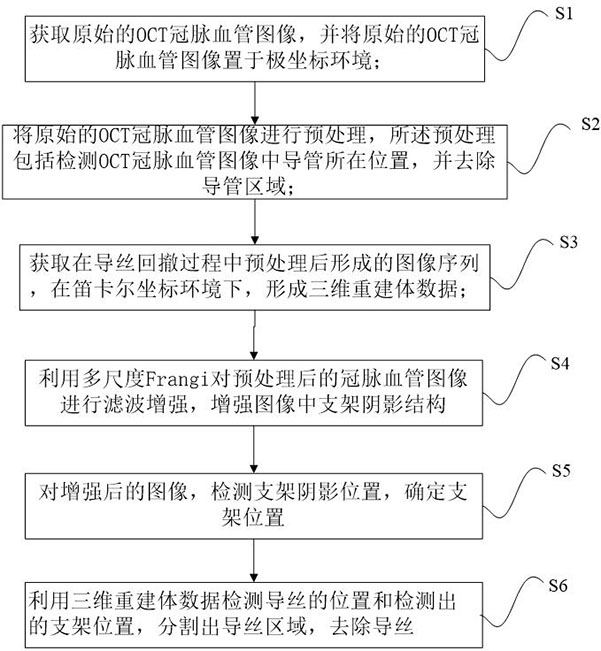
[0062] like figure 1 As shown, the method for detecting coronary stents based on OCT images provided by an embodiment of the present invention includes the following steps

[0063] S1. Obtain an original OCT coronary vessel image, and place the original OCT coronary vessel image in a polar coordinate environment;

[0064] S2, preprocessing the original OCT coronary vessel image, the preprocessing includes detecting the position of the catheter in the OCT coronary vessel image, and removing the catheter area;

[0065] S3, acquiring the image sequence formed after preprocessing in the guide wire retraction process, and forming three-dimensional reconstructed volume data in a Cartesian coordinate environment;

[0066] S4. Use multi-scale Frangi to filter and enhance the preprocessed coronary vessel image to enhance the shadow structure of the sten...


PUM


Abstract

The invention discloses a coronary artery intravascular stent detection method and system based on an OCT image, and the method comprises the steps: obtaining an original OCT coronary artery image, and placing the image in a polar coordinate environment; the original OCT coronary blood vessel image is preprocessed, and preprocessing comprises the steps that the position of a catheter in the OCT coronary blood vessel image is detected, and a catheter area is removed; acquiring an image sequence formed in a guide wire withdrawing process, and forming three-dimensional reconstruction body data in a Cartesian coordinate environment; performing filtering enhancement on the preprocessed coronary blood vessel image by using multi-scale Frangi, and enhancing a stent shadow structure in the image; detecting the shadow position of the bracket for the enhanced image, and determining the position of the bracket; and detecting the position of the guide wire and the detected position of the stent by using the three-dimensional reconstruction volume data, segmenting a guide wire region, and removing the guide wire. According to the method, after multi-scale Frangi filtering enhancement is utilized, bracket shadow position detection and guide wire removal are carried out, the noise influence is reduced, and a high-fidelity image is obtained.

Description

technical field [0001] The invention relates to the technical field of medical image processing, and in particular, to a method and a detection system for detecting a coronary intravascular stent based on an OCT image. Background technique [0002] Globally, cardiovascular and cerebrovascular diseases have become one of the major diseases threatening human health, and coronary atherosclerosis is the main cause of cardiovascular and cerebrovascular diseases. At present, coronary stent interventional therapy has become a main treatment plan for coronary atherosclerotic disease because of its small trauma and good effect. During treatment, stents are placed inside the coronary arteries through treatment to reduce the chance of restenosis and thrombosis. [0003] Optical coherence tomography (optical coherence tomography, OCT) is the latest intravascular imaging technology for the diagnosis of cardiovascular and cerebrovascular diseases. OCT utilizes near-infrared light and is...

Claims


Application Information

Patent Timeline
Patent Type & Authority Applications(China)
IPC IPC(8): G06T7/00G06T7/73G06T7/10G06T5/00
CPCG06T7/0012G06T7/73G06T7/10G06T2207/10101G06T2207/30021G06T2207/30052G06T2207/30101G06T5/94
Inventor 武西宁赵士勇
Owner HORIMED TECH CO LTD
Learn More