Method and reagent for identifying antibody combined with mutant antigen
A mutation and antigen technology, applied in the field of immune detection, can solve the problems of high incidence
- Summary
- Abstract
- Description
- Claims
- Application Information
AI Technical Summary
Problems solved by technology
Method used
Image
Examples
Embodiment 1
[0047] Example 1 Identification of mutants by colloidal gold platform
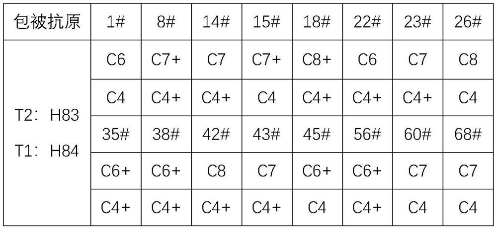
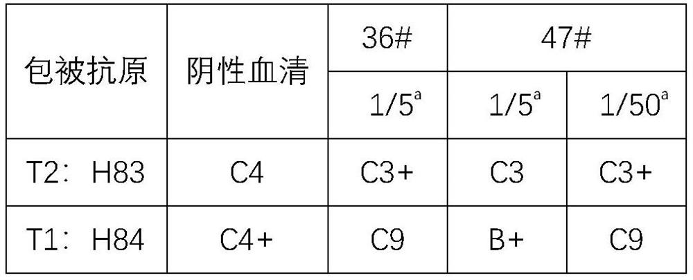
[0048] The amino acid sequence of H83 antigen is shown in SEQ ID NO: 1, the amino acid sequence of H84 antigen is shown in SEQ ID NO: 2, Ab13 antibody is an antibody that binds to a reference antigen (such as H83 antigen); Ab13 antibody, human ACE2 can be purchased from Feipeng Biological; other reagents and materials are commercially available.
[0049] 1. Mark
[0050] (1) Preparation of colloidal gold: adopt the traditional sodium citrate reduction method, first heat the chloroauric acid solution to boiling, quickly add a certain proportion of trisodium citrate solution, stir evenly, and wait until the color of the solution becomes wine red and no longer changes Stop heating, cool to room temperature to obtain a colloidal gold solution with a concentration of 4 / 10,000;
[0051] (2) Labeling: Add 0.2M K to the colloidal gold solution 2 CO 3 The solution is adjusted to pH 6.0-7.5;
[0052] (3) Centri...
Embodiment 2
[0069] Example 2 Identification of mutants by colored microspheres
[0070] 1. Mark
[0071] Take colored microspheres, after 300W ultrasound, add 0.1ml of latex particles to 0.9ml of 100mM MES, vortex and mix; centrifuge at 15000rmp for 15min, then remove the supernatant; add 1.0ml of 100mM MES for ultrasound, add appropriate amount of MES and NHS to activate the microspheres 10min; centrifuge at 15000rmp for 15min, then remove the supernatant; add 1.0ml of 100mM MES for ultrasound, add an appropriate amount of ACE2, vortex overnight at 37°C; centrifuge at 15,000rmp for 15min, remove the supernatant, add BSA to block, sonicate, and react at 37°C for 4h; 15000rmp for 15min Centrifuge, remove the supernatant, wash, and sonicate; centrifuge at 15,000 rmp for 15 min, remove the supernatant and resuspend; dilute the resuspended concentrate, spread it on glass cellulose membrane, and then put it in a freeze dryer for lyophilization (1-2h) Or put it in a drying room at 37°C to dry ...
Embodiment 3
[0091] Example 3 Identification of mutants by colored microspheres
[0092] The H85 antigen amino acid sequence is shown in SEQ ID NO:3. The H85 antigen was coated on the T1 detection line, the H83 antigen was coated on the T2 detection line, and other labeling, coating, and detection processes were the same as those in Example 2. When the test sample is negative serum, the color development of the T1 and T2 detection lines is equivalent; when the detection sample contains an antibody that binds to the non-mutated antigen including the 456-position phenylalanine of the spike protein, the color development of the T2 detection line is weakened. The color of the T1 detection line is stronger than that of the T2 by at least 2 color cards; when the test sample contains an antibody that binds to the 456-position alanine of the spike protein, the color development of the T1 detection line is weakened and almost disappears, and the color of the T2 detection line is developed Stronger...
PUM
 Abstract
Description
Claims
Application Information
 - R&D
- Intellectual Property
- Life Sciences
- Materials
- Tech Scout
- Unparalleled Data Quality
- Higher Quality Content
- 60% Fewer Hallucinations
Browse by: Latest US Patents, China's latest patents, Technical Efficacy Thesaurus, Application Domain, Technology Topic, Popular Technical Reports.
© 2025 PatSnap. All rights reserved.Legal|Privacy policy|Modern Slavery Act Transparency Statement|Sitemap|About US| Contact US: help@patsnap.com


