Method of biological tissue optical and ultrasonic collection and tomographic imaging and its device

A biological tissue and tomographic imaging technology, applied in the field of imaging, can solve the problems of inability to make benign or malignant judgments, poor imaging contrast, etc., and achieve the effects of simple structure, easy assembly and convenient operation.

Inactive Publication Date: 2004-12-22
SOUTH CHINA NORMAL UNIVERSITY
View PDF0 Cites 30 Cited by
  • Summary
  • Abstract
  • Description
  • Claims
  • Application Information

AI Technical Summary

Problems solved by technology

The advantage of the B-ultrasound instrument is multi-probe imaging (the linear array probe contains hundreds of probe units), fast im...

Method used


Image

  • Method of biological tissue optical and ultrasonic collection and tomographic imaging and its device
  • Method of biological tissue optical and ultrasonic collection and tomographic imaging and its device
  • Method of biological tissue optical and ultrasonic collection and tomographic imaging and its device

Examples


Embodiment Construction

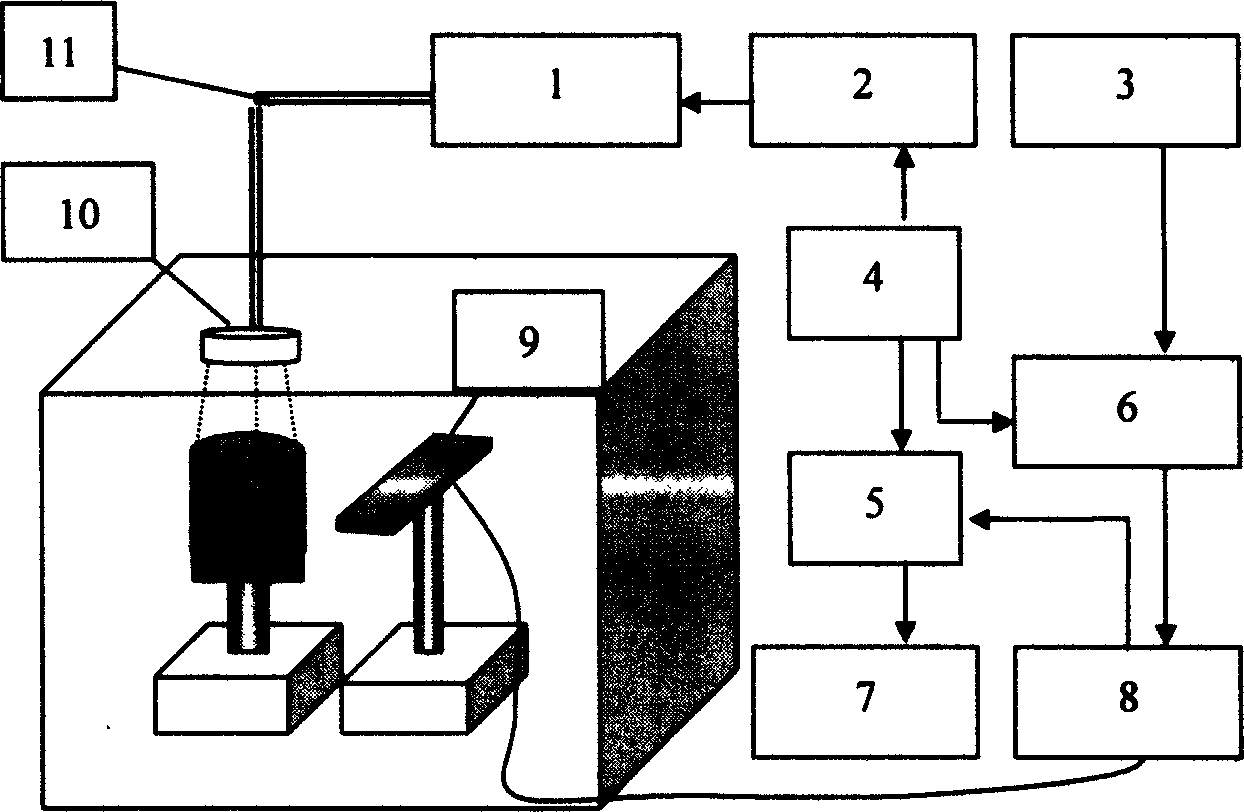
[0028] The device structure of the present invention is as attached figure 1 As shown, the device of the present invention is mainly composed of a laser 1, a trigger delay circuit 2, a 100KHz external trigger source circuit 3, a 30Hz external trigger circuit 4, a high-speed data acquisition card 5, an external trigger circuit selection switch 6, a computer 7, and a phase control focusing circuit 8. Composed of multi-element ultrasonic array detector 9, collimating lens 10, and optical fiber 11; among them, multi-element ultrasonic array detector 9 is electrically connected with phase control focusing circuit 8 to form a multi-element ultrasonic array; 30Hz external trigger circuit 4, trigger delay circuit 2 1, the laser 1 is electrically connected; the multi-element ultrasonic array, the data acquisition card 5, and the computer 7 are electrically connected sequentially; the 30Hz external trigger circuit 4 is simultaneously connected with the high-speed data acquisition card 5,...


PUM

PropertyMeasurementUnit
Wavelengthaaaaaaaaaa

Abstract

A method for optically and ultrasonically acquiring the information about biological tissue and chromatographic imaging includes ultrasonically scanning the biologic tissue to find out possible lesion position, emitting the laser pulses and ultasonic waves to the tissue at lesion position to obtain photoultrasonic and reflected ultrasonic signals, receiving the ultrasonic echo and photo-acoustical signals, data processing by computer, and straight projection for chromatographic imaging. Its equipment is composed of laser, multi-element ultrasonic array, externally triggered signal source, data acquisition card, delay circuit, and computer.

Description

technical field [0001] The invention belongs to imaging technology, and more specifically relates to a method and device for optical and ultrasonic acquisition and tomographic imaging of biological tissue. technical background [0002] The application of photoacoustic methods in medicine, especially the diagnosis of breast cancer, has become a hot spot in current research. Most researchers believe that this is a promising diagnostic method, especially for the diagnosis of early breast cancer, because breast cancer The main component of the tissue is fat. The capillaries around the early cancerous tissue are very rich, and the blood oxygen content in the blood vessels is high, which leads to a large difference in the optical absorption characteristics between it and the surrounding fat tissue. Experiments have shown that the light absorption of breast cancer tissue It is more than 5 times higher than that of normal tissue, and the tomographic imaging...

Claims


Application Information

Patent Timeline
IPC IPC(8): A61B8/00A61B8/13
Inventor 邢达曾亚光尹邦政
Owner SOUTH CHINA NORMAL UNIVERSITY
Why Eureka
Try Eureka