
Bionic stent generating method based on CT picture

A CT image and bionic technology, applied in image data processing, special data processing applications, 3D modeling, etc., can solve the problems of uncontrollable internal micropores and inability to completely match bone tissue defects

Inactive Publication Date: 2005-04-27
SHANGHAI UNIV
View PDF0 Cites 23 Cited by
  • Summary
  • Abstract
  • Description
  • Claims
  • Application Information

AI Technical Summary

Problems solved by technology

[0003] The manufacturing method of the above-mentioned bionic scaffold cannot completely match the bone tissue defect in terms of macrostructure, and is affected by materials and environmental factors in microstructure, so the generated internal micropores are uncontrollable

Method used


Image

  • Bionic stent generating method based on CT picture
  • Bionic stent generating method based on CT picture
  • Bionic stent generating method based on CT picture

Examples


Embodiment Construction

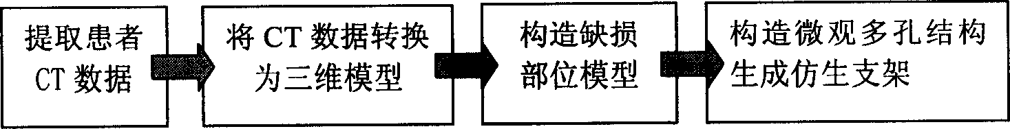
[0027] A preferred embodiment of the present invention is described in detail as follows: In this embodiment, according to the cranial CT image data of patients with bone tissue defects provided by the hospital, a bionic scaffold model with an internal microporous structure is obtained through computer processing. The method is: according to the CT image of the patient's bone, it is first converted into a three-dimensional bone model, and then a three-dimensional model of the bone tissue defect is established from the bone model, and then a bionic scaffold model with an internal microporous structure suitable for the growth of bone seed cells is constructed . The microporous structure will meet the requirements of seed cell growth, that is, the porosity will reach 80%-90%, the pore diameter will be between 0.2-0.5mm (the pore size is controllable), and the connectivity of the pores will be guaranteed.

[0028] see figure 1 , the steps of this method are:

[0029] (1) First o...


PUM


Abstract

The method of creating bionic stent based on CT image includes: converting the hospital provided CT image data of the patient into 3D model; constituting the 3D model of bone tissue deficiency position; creating non-homogeneous porous geometrical description model based on tissue engineering requirement and finally creating 3D bionic stent model with internal micro structure, suitable for the growth of bone seed cell and used for repairing bone tissue deficiency.

Description

Technical field: [0001] This patent relates to a bionic scaffold generation method for bone tissue defect repair in the field of human tissue engineering. In particular, a bionic scaffold generation method based on CT images. Background technique: [0002] For the widely used CT image data in medicine, it has been reported abroad that it can be converted into a three-dimensional model, and according to the nature of CT images with different pixel gray levels for different tissues and lesions, it can be observed, judged, analyzed, and positioned. Tissue or lesion location, this is only in the macroscopic sense, and there is no report on the modeling method of the microscopic bionic medical model, that is, the three-dimensional bionic scaffold model with internal microscopic porous structure. On the other hand, for the repair of bone tissue defects, the required bionic scaffold model can only be obtained manually and by physical and chemical methods (ie, fiber bonding, gas fo...

Claims


Application Information

Patent Timeline
Patent Type & Authority Applications(China)
IPC IPC(8): G06F17/50G06T17/00
Inventor 戴春祥李莉敏胡庆夕吴懋亮陆齐方明伦谭月梅
Owner SHANGHAI UNIV
Learn More