Ultrasonic imaging apparatus

A technology of ultrasonic imaging and ultrasonic image, which is applied in ultrasonic/acoustic/infrasonic diagnosis, acoustic diagnosis, infrasonic diagnosis, etc. It can solve the problems of unnatural data comparison, inability to obtain reliable morphological comparison, unintuitive and other problems, and achieve reliable comparison Effect

Inactive Publication Date: 2006-06-14
GE MEDICAL SYST GLOBAL TECH CO LLC
View PDF1 Cites 10 Cited by
  • Summary
  • Abstract
  • Description
  • Claims
  • Application Information

AI Technical Summary

Problems solved by technology

[0006] According to the prior art as already explained above, data comparison using the measurement graph can be performed for numerical data, however, the data comparison is neither natural nor intuitive
On the other hand, when the visual comparison of the anatomical morphology set by the measurement patterns is performed for each image by merging multiple ultrasound images of the imaging locations in a kinetic etiological order, the morphological comparison between the measurement patterns may become too much. ambiguous so that reliable morphological comparisons cannot be obtained

Method used


Image

  • Ultrasonic imaging apparatus
  • Ultrasonic imaging apparatus
  • Ultrasonic imaging apparatus

Examples


Embodiment Construction

[0048] Hereinafter, the best mode for carrying out the ultrasonic imaging apparatus according to the present invention will be described in detail with reference to the accompanying drawings. It should be noted here that the illustrated embodiments are not intended to limit the invention.

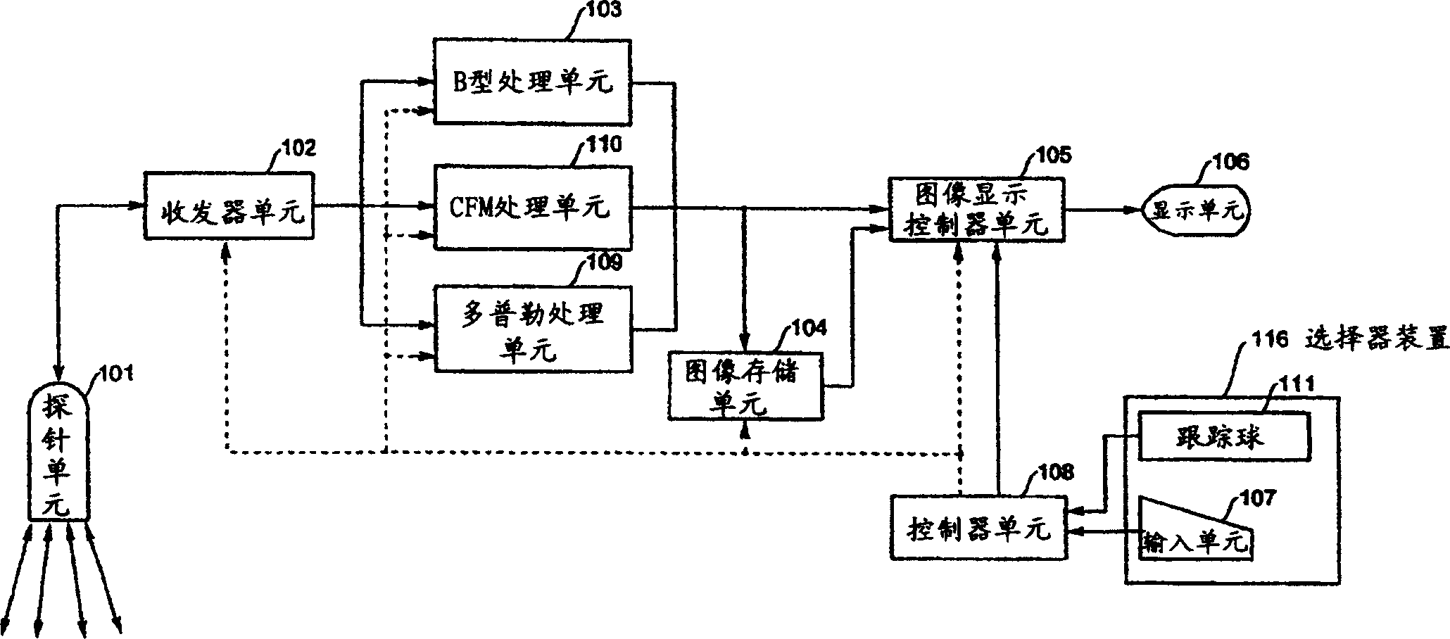
[0049] figure 1 A schematic block diagram indicating an overview of an ultrasound imaging apparatus according to a preferred embodiment of the present invention is shown. The ultrasonic imaging device has a probe 101, a transceiver unit 102, a B-type processing unit 103, a Doppler processing unit 109, a color flow mapping (CFM) processing unit 110, an image storage unit 104, an image display control The device unit 105, the display unit 106, the selector device 116 and the controller unit 108. It is worth noting that the following will describe the figure 1 and figure 2 In , signal lines for transmitting image information or selection information are indicated by solid lines, while sig...


PUM


Abstract

The present invention provides an ultrasound imaging device which allows to carry out a morphological comparison of measurement patterns set within a plurality of ultrasound images in a reliable and easier manner. The measurement pattern storage device (250) stores the circle indicating the size of the inner wall of the left ventricle, and is designated in the B-mode image of the diastolic period of the left ventricle, and the measurement pattern reproduction device (240) reproduces the circle indicating the systole period of the left ventricle in the B-mode image, so that A morphological comparison of the left ventricular inner wall in systole and diastole can be done in an easier and very reliable manner with the Numerical data are obtained together in order to provide an appropriate determination of the numerical data.

Description

technical field [0001] The present invention relates to an ultrasound imaging apparatus that displays an ultrasound image of a subject, and displays a measurement pattern in the displayed ultrasound image based on measurement pattern information on the position and shape of the input measurement pattern, so as to implement the measurement of the ultrasound image Image measurement. Background technique [0002] Recently, in ultrasonic imaging apparatuses, ultrasonic images as generally represented by "B" type images have fine grains. Furthermore, not only morphological examinations can be performed in real time, but also measurements and examinations of organ anatomy can be performed on the basis of quantitative information derived therefrom. [0003] In quantitative inspection, a measurement pattern including such as a trajectory is loaded into an ultrasound image, and the pattern is repositioned to a target position by using a pointing device such as a trackball for image ...

Claims


Application Information

Patent Timeline
Patent Type & Authority Applications(China)
IPC IPC(8): A61B8/00
CPCA61B5/1075A61B8/06A61B8/08A61B8/0883A61B8/13A61B8/14A61B8/463A61B8/467A61B8/488A61B8/5238G01S7/52073G01S7/52074G01S7/52084A61B8/00
Inventor 伊藤真由美阿部弥生藤原千织八幡努
Owner GE MEDICAL SYST GLOBAL TECH CO LLC
Why Eureka
Try Eureka