Eureka AIR delivers breakthrough ideas for toughest innovation challenges, trusted by R&D personnel around the world.

Method for distinguishing epithelial carcinoma property by single cell Raman spectrum

A technology of Raman spectroscopy and epithelial cells, which is applied in Raman scattering, biochemical equipment and methods, measurement/testing of microorganisms, etc., which can solve the problems of spectral differences, loss of biological activity of tissue samples, detection technology is difficult to avoid interference, etc question

Inactive Publication Date: 2006-07-19
SHANGHAI INST OF OPTICS & FINE MECHANICS CHINESE ACAD OF SCI
View PDF0 Cites 7 Cited by
  • Summary
  • Abstract
  • Description
  • Claims
  • Application Information

AI Technical Summary

Problems solved by technology

[0009] (2) The tissue sample has lost its biological activity, and its spectrum is different from that of the tissue when it is in a living state inside the human body;
Since 85% of cancers originate from epithelial cell variation, epithelial cells should be the main detection target, while blood cells in subcutaneous tissue are the source of strong Raman signals, and it is difficult to avoid interference with tissue-level detection techniques

Method used


Image

  • Method for distinguishing epithelial carcinoma property by single cell Raman spectrum
  • Method for distinguishing epithelial carcinoma property by single cell Raman spectrum
  • Method for distinguishing epithelial carcinoma property by single cell Raman spectrum

Examples


preparation example Construction

[0027] (1) Preparation of single-cell samples and maintenance of biological activity

[0028] The present invention can share the sample source with the current routine medical pathological detection in the hospital. Usually, in colorectal tumor surgery, in addition to tumor tissue removal, a small amount of normal mucosal tissue near the lesion site is also removed, and the latter is used as a normal control in medical pathological testing.

[0029] The sample processing steps of the present invention are as follows: Immediately after the operation of the colorectal tumor patient, about 0.5 cm each was cut from the surgically removed fresh tumor tissue and normal mucosal tissue under aseptic conditions. 3 , the latter was taken from the mucosa more than 10cm away from the tumor. Place the collected tissue in D-Hanks balanced salt solution containing penicillin 300U / ml and streptomycin 300μg / ml for 20 minutes, and then cut it into a size less than 1mm 3 The fragments of the ...

Embodiment 1

[0077] Zhu X X, male, 52 years old, pathologically diagnosed as sigmoid colon ulcer type adenocarcinoma, highly to moderately differentiated, adopts the method of the present invention to carry out double-blind detection. The doctor provided single-cell samples from the lesion and non-lesion sites, and the spectral detection correctly identified 17 of the 20 normal cells and 17 of the 20 cancer cells. The results showed that the patient was indeed suffering from cancer. Figure 5 For embodiment 1 diagnosis result.

Embodiment 2

[0079] Shen X X, male, 64 years old, pathologically diagnosed as rectal infiltrative ulcer type adenocarcinoma, well differentiated, adopting the method of the present invention to carry out double-blind detection. The doctor provided single-cell samples from the lesion and non-lesion sites. The spectral detection correctly identified all 20 normal cells and 16 of the 20 cancer cells. The results showed that the patient was indeed suffering from cancer. Figure 6 It is embodiment 2 diagnosis result.


PUM


Abstract

The invention relates to a method for using the single-cell Laman spectrum to quote the epithelial cell cancer nature. It comprises: preparing for the human focusing position single-cell sample and holding the biology activity, measuring the single live cell Laman spectrum near to the human physiological condition, quoting weather the cell is cancer or normal and the applied program of the single-cell Laman spectrum diagnose identifying model.

Description

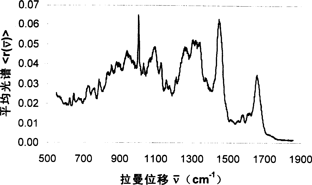
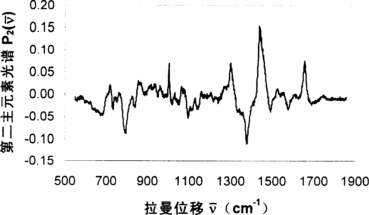
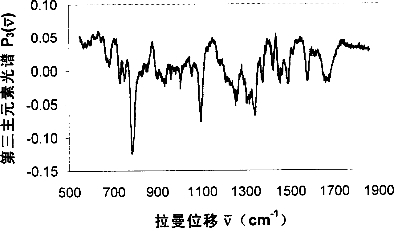
technical field [0001] The invention relates to a method for distinguishing the nature of epithelial cell cancer by single-cell Raman spectroscopy, which can be used for the diagnosis of epithelial cell canceration, including colorectal cancer, bladder cancer, esophageal cancer, skin cancer, oral cancer and other cancers. Background technique [0002] At present, medical pathology detection (pathology) is the standard method for the diagnosis of cancer. Its operation process is to fix the fresh tissue of the surgically resected lesion with formalin and embed it in paraffin. Pathologists observe under a microscope and draw conclusions based on experience. Usually, formalin fixation takes 3-6 hours, and section preparation and staining take 24 hours, so it takes a long time to reach the stage of pathological analysis. What's more, the pathological conclusion obtained from the slice observation is the subjective judgment of pathologists based on experience, and there is a cert...

Claims


Application Information

Patent Timeline
IPC IPC(8): G01N21/65C12Q1/02
Inventor 陈昆覃业军魏青孙孟红邓见辽周晓燕
Owner SHANGHAI INST OF OPTICS & FINE MECHANICS CHINESE ACAD OF SCI
Why Eureka
Eureka Blog
Learn More