3D refractive index tomography and structured illumination microscopy system using wavefront shaper and method thereof

a technology of refractive index and structured illumination, applied in the direction of instruments, optical elements, fluorescence/phosphorescence, etc., can solve the problems of difficult to distinguish a specific protein, invasive process of dying a cell to express fluorescent proteins, and inability to measure the cell, etc., to achieve high resolution

Active Publication Date: 2018-09-25
KOREA ADVANCED INST OF SCI & TECH +1
View PDF7 Cites 1 Cited by
  • Summary
  • Abstract
  • Description
  • Claims
  • Application Information

AI Technical Summary

Benefits of technology

[0035]The fluorescence image unit may make the plane wave incident on the sample with a pattern, may obtain a plurality of fluorescence images by using patterns controlled by adjusting a phase between plan

Problems solved by technology

However, it is difficult to distinguish a specific protein because the optical diffraction tomography is not a specific measure at the molecular level.
However,

Method used


Image

  • 3D refractive index tomography and structured illumination microscopy system using wavefront shaper and method thereof
  • 3D refractive index tomography and structured illumination microscopy system using wavefront shaper and method thereof
  • 3D refractive index tomography and structured illumination microscopy system using wavefront shaper and method thereof

Examples


Embodiment Construction

[0043]Hereinafter, embodiments of the inventive concept will be described with reference to accompanying drawings. However, embodiments to be described may be modified in the different forms, and the scope and spirit of the inventive concept is not limited by the embodiments to be described below. In addition, various embodiments are provided to describe this disclosure more fully to those skilled in the art. For a clear description, forms, sizes, and the like of elements may be exaggerated in a drawing.

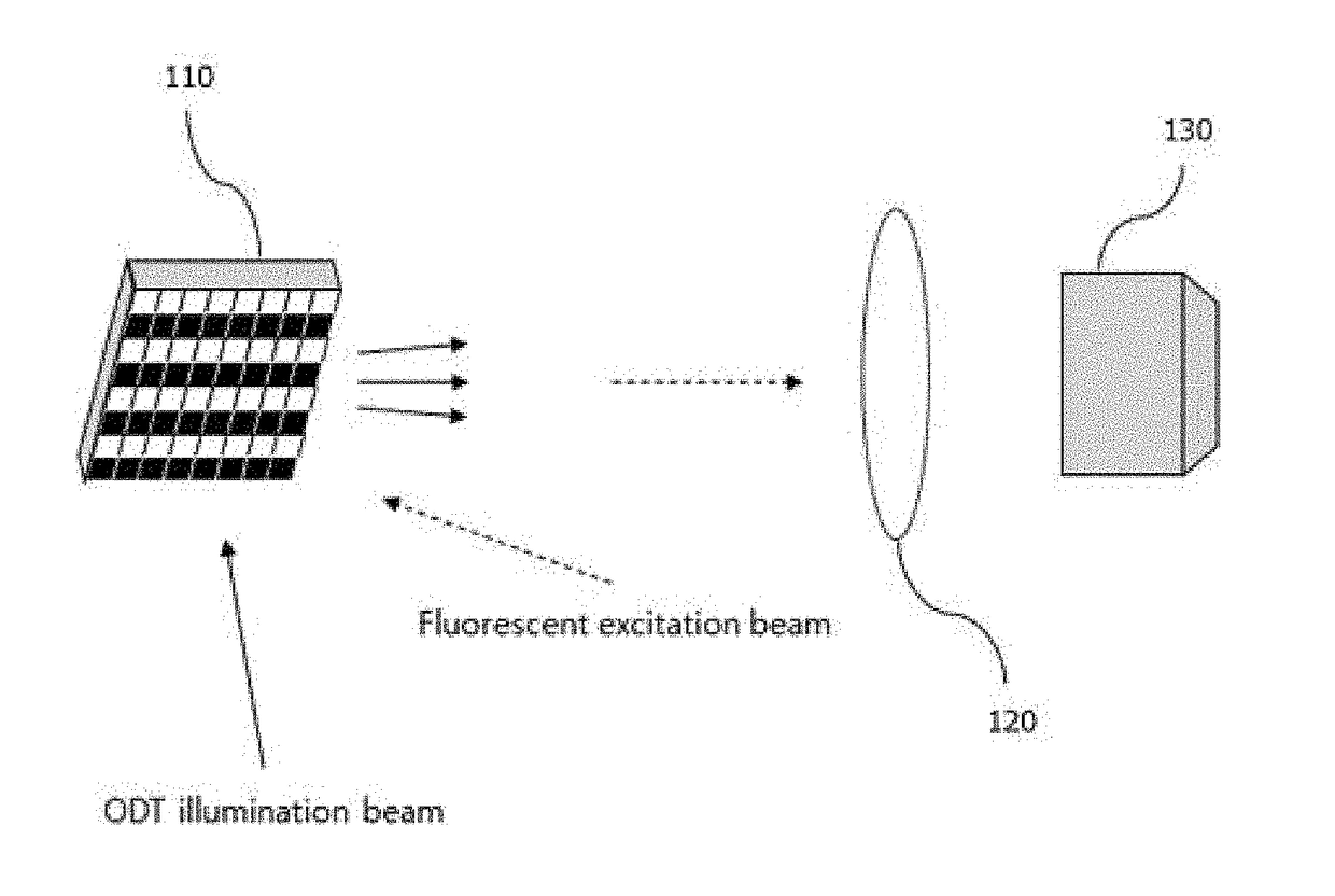
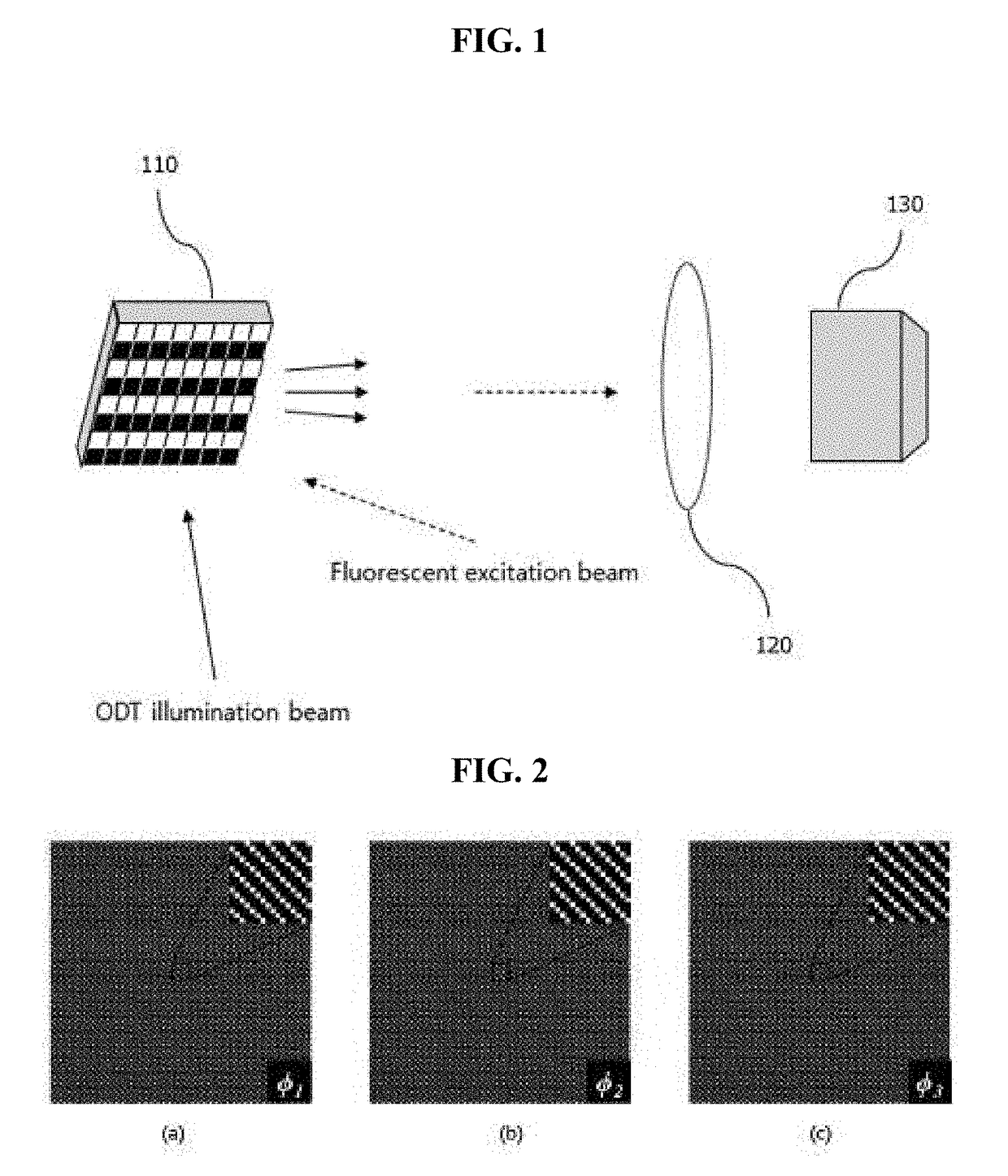
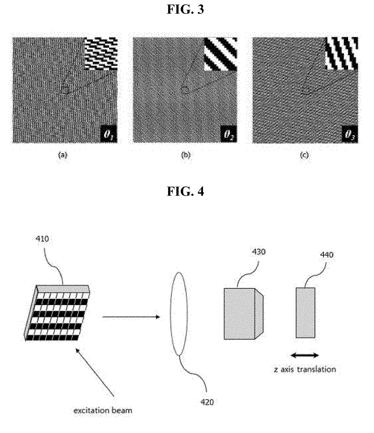
[0044]To three-dimensionally analyze an inner structure of a cell and to measure the change in a structure in real time may be a technology that greatly contributes to the biological and pathological studies.

[0045]Embodiments to be described below provide a system and method that implements both a 3D refractive index tomography and a 3D structured illumination microscopy by using a wavefront shaper.

[0046]Embodiments may provide a technology that is capable of simultaneously measuring...


PUM


Abstract

An ultra-high-speed 3D refractive index tomography and structured illumination microscopy system using a wavefront shaper and a method using the same are provided. A method of using an ultra-high-speed 3D refractive index tomography and structured illumination microscopy system that utilizes a wavefront shaper includes adjusting an irradiation angle of a plane wave incident on a sample by using the wavefront shaper, measuring a 2D optical field, which passes through the sample, based on the irradiation angle of the plane wave, and obtaining a 3D refractive index image from information of the measured 2D optical field by using an optical diffraction tomography or a filtered back projection algorithm.

Description

CROSS-REFERENCE TO RELATED APPLICATIONS[0001]A claim for priority under 35 U.S.C.§ 119 is made to Korean Patent Application No. 10-2016-0072304 filed Jun. 10, 2016, in the Korean Intellectual Property Office, the entire contents of which are hereby incorporated by reference.BACKGROUND[0002]Embodiments of the inventive concepts described herein relate to an ultra-high-speed 3-dimensional (3D) refractive index tomography and structured illumination microscopy system using a wavefront shaper and a method using the same, and more particularly, to a technology that simultaneously measures a 3D high resolution fluorescence image and a 3D refractive index stereoscopic image of a cell by using a single wavefront shaper.[0003]Nowadays, methods used to optically measure an inner structure of a cell are a fluorescence image method that expresses a fluorescent protein in a specific structure of a cell and measures a fluorescence signal and a 3D refractive index tomography that measures a 3D ref...

Claims


Application Information

Patent Timeline
IPC IPC(8): G02B21/36G03H1/00G02B27/46G01N21/45G02B26/08G02B21/16G01N21/41G01N21/64G01N21/17
CPCG02B21/367G01N21/4133G01N21/45G01N21/6458G01N21/6486G02B26/0833G02B27/46G03H1/0005G02B21/16G03H2001/005G01N2021/1787G01N2201/0635G01N21/455G01N21/6456G01N2021/6495G01N21/453G02B6/32G02B6/3516
Inventor PARK, YONGKEUNSHIN, SEUNGWOOPARK, GWANG SIK
Owner KOREA ADVANCED INST OF SCI & TECH
Why Eureka
Try Eureka