Method and apparatus for recording a magnetic resonance dataset of at least one foreign body in a patient

a technology of magnetic resonance dataset and foreign body, which is applied in the field of method and apparatus for recording a magnetic resonance dataset of at least one foreign body in a patient, can solve the problems of loss of structural integrity and/or functionality of the foreign body, failure of cemented implants, and failure of polymeric fastenings, glues and cements, etc., to achieve fast signal decay, simplify segmentation, and facilitate segmentation

Active Publication Date: 2019-04-23
THE JOHN HOPKINS UNIV SCHOOL OF MEDICINE +1
View PDF15 Cites 0 Cited by
  • Summary
  • Abstract
  • Description
  • Claims
  • Application Information

AI Technical Summary

Benefits of technology

[0017]In a preferred specific embodiment of magnetic resonance measurement in order to be able to perform a segmentation of the foreign body in a simplified manner, as part of the magnetic resonance sequence, a saturation pulse having a duration of at least 25 ms, preferably at least 30 ms, is radiated before the excitation pulse, so that the signal from proton spins lying outside the foreign body and exceeding the threshold value for the T2* relaxation time is saturated. By virtue of the fact that in the region of the foreign body, due to the short T2 and T2* times, an extremely fast signal decay takes place, the relevant spins do not reach saturation but are only somewhat excited, whereupon the excitation quickly decays again, and such like. This means that after the saturation pulse the measurement can be made after just a short waiting time for relaxation of the slight excitation, whereas the spins with long T2 or T2* relaxation times are in saturation and deliver no signal at all. In this embodiment, it is therefore possible for only the magnetic resonance signal of the foreign body will be measured, which may simplify segmentation.
[0018]It should also be noted that in the embodiment with a measurement at various echo times, a similar image can also be generated by making a second measurement at a significantly later second echo time, the magnetic resonance signals of the foreign body having at the time of the second measurement long since decayed. If one then removes from the magnetic resonance image of the magnetic resonance dataset produced in the first measurement at the ultra-short echo time, the magnetic resonance image of the magnetic resonance dataset recorded at the second echo time in the second measurement, a magnetic resonance image is also obtained that contains only the portions of the foreign body, so that here, too, segmentation is easy.
[0020]With such evaluation variables, in particular T2* relaxation times, which are available as a map, thus for each pixel, a large number of automatic and manual further evaluation procedures can be carried out. Thus, for example, it can be seen from a T2* map where higher T2* times than expected in the foreign body occur, which is an indication of a crack or another structural integrity defect. Thus deviations in these evaluation variables can show damage to the foreign body or an infection. It is thus possible to identify even small deviations, so that a problem can be identified even at an early stage of damage, infection or the like, so that appropriate remedial procedures can be implemented. Thus, generally speaking, it can be provided that at least one item of structural integrity information is determined by evaluating the evaluation variables in the foreign body, in particular by comparing with target values and / or by analyzing the distribution of the evaluation variables. If, for example, significantly longer T2* values occur in an area of the foreign body, it can be assumed that either the protons are not so strongly bound or that free water protons are present. In both cases, this may indicate damage, for example cracks or the like, to the foreign body, for example an implant.

Problems solved by technology

If such a foreign body is introduced into a patient or produced in the patient, complications often occur, for example rejection reactions by the body, infections or mechanical faults leading to the loss of the structural integrity and / or the functionality of the foreign body.
For example, cemented implants may not integrate correctly in the relevant bones or may fail by becoming detached after successful implantation.
Polymeric fastenings, glues and cements may become defective due to degradation over time, for example crack formation and such like.
Biodegradable devices such as retaining screws may fail in their intended function due to inflammatory responses in patients or to breakage.
For application cases of this kind, in which a non-invasive evaluation of the foreign body, for example an implant, is desirable, imaging modalities such as computed tomography and ultrasound are less suitable, because image artifacts often occur, the contrast is unsuitable and inflammations or such like do not become visible.
In principle, magnetic resonance imaging is therefore an obvious choice as an imaging modality, since it provides high signal and contrast resolution of soft tissues and bones, but it has not been capable of visualizing the integrity of foreign bodies, for example solid implants and fastening components.
Magnetic resonance imaging normally cannot be used because the magnetic resonance signal has already decayed before the current magnetic resonance sequences could measure it.
In addition, pronounced image artifacts can occur, because metallic foreign bodies, in particular, can distort the fields of the magnetic resonance scanner that are necessary for measurement, due to their susceptibility effects.
While magnetic resonance sequences that reduce these artifacts and permit imaging of the tissue around the foreign body have become known, no method for imaging the foreign bodies themselves, which is suitable for extensive clinical practice, has yet become known.

Method used


Image

  • Method and apparatus for recording a magnetic resonance dataset of at least one foreign body in a patient
  • Method and apparatus for recording a magnetic resonance dataset of at least one foreign body in a patient

Examples


Embodiment Construction

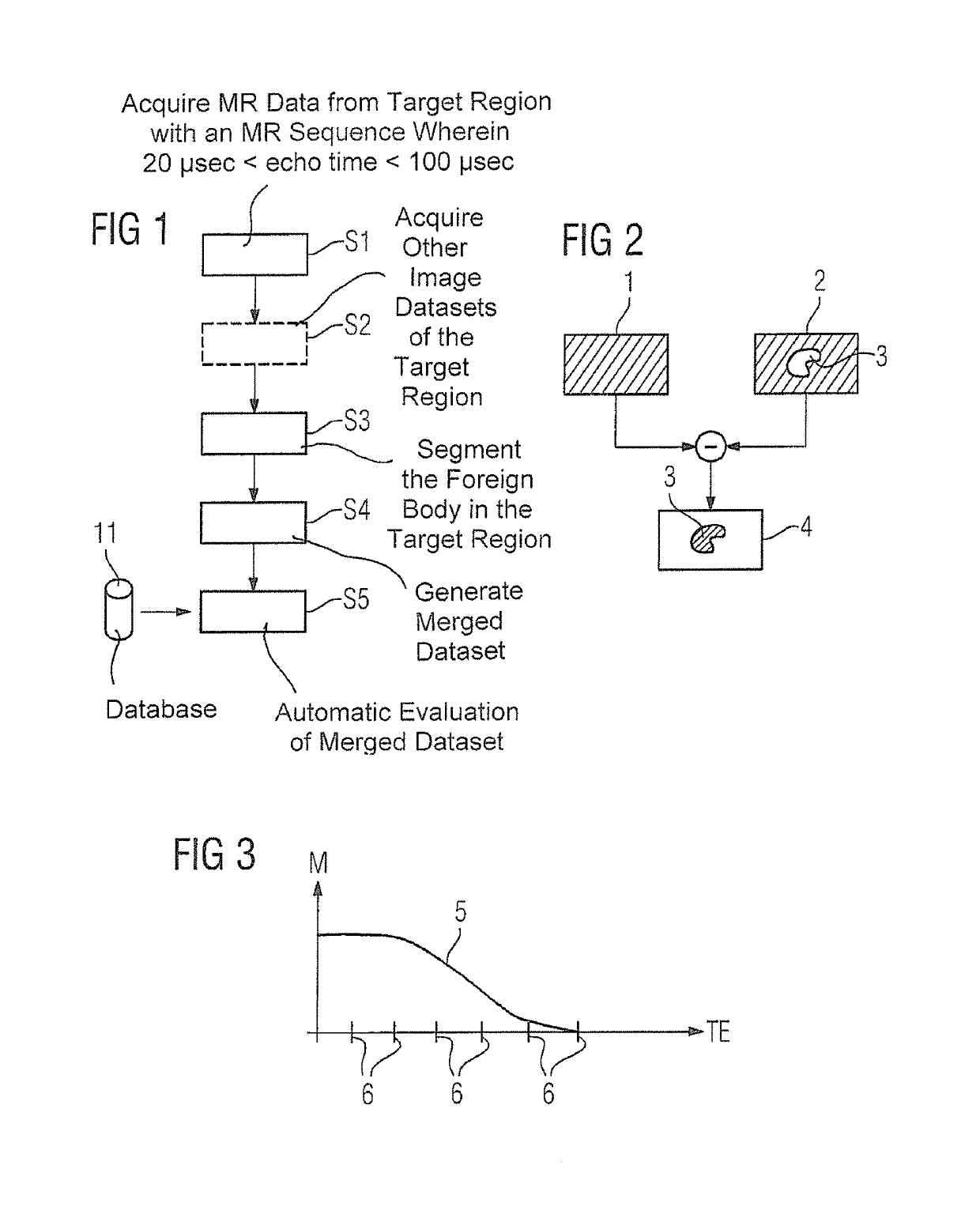
[0034]FIG. 1 IS a flowchart of an exemplary embodiment of the inventive method which is for recording and evaluating magnetic resonance data of a foreign body in a target region of a patient, in the specific example of an implant. Here, not only is magnetic resonance data of the implant itself, for example of a replacement joint, to be recorded, but also an automatic evaluation carried out with regard to the functionality and structural integrity of the implant and influence on its surroundings. Of course, this description also applies to other foreign bodies, for example cements, fastenings, glues, screws, and other medical devices.

[0035]In a step S1 the magnetic resonance data are recorded with a magnetic resonance scanner, a target region of the patient containing the implant being selected as the coverage area. A magnetic resonance sequence is used which permits ultra-short echo times, here in the range from 20 μs to 100 μs, i.e. the first measurement of magnetic resonance data ...


PUM


Abstract

In a method and apparatus for recording a magnetic resonance dataset of at least one foreign body in a target region of a patient, a magnetic resonance sequence having an ultra-short echo time, which is less than 500 μs is used for recording the magnetic resonance data.

Description

BACKGROUND OF THE INVENTION[0001]Field of the Invention[0002]The present invention concerns a method for recording a magnetic resonance dataset of at least one foreign body in a target region of a patient, as well as and to a magnetic resonance apparatus designed to implement such a method.[0003]Description of the Prior Art[0004]In surgical procedures, foreign bodies may be introduced into a patient. While implants are a classic example of such foreign bodies, glues, fillings, foreign-material fastenings, cementations and other, possibly functional, medical devices must also be mentioned. If such a foreign body is introduced into a patient or produced in the patient, complications often occur, for example rejection reactions by the body, infections or mechanical faults leading to the loss of the structural integrity and / or the functionality of the foreign body. For example, cemented implants may not integrate correctly in the relevant bones or may fail by becoming detached after suc...

Claims


Application Information

Patent Timeline
Patent Type & Authority Patents(United States)
IPC IPC(8): G01R33/48G01R33/50G01R33/56G01R33/565
CPCG01R33/4816G01R33/50G01R33/5608G01R33/56536
Inventor FRITZ, JANGRODZKI, DAVIDMEYER, HEIKONITTKA, MATHIAS
Owner THE JOHN HOPKINS UNIV SCHOOL OF MEDICINE
Why Eureka
Try Eureka