
Methods and apparatus for the on-line and real time acquisition and analysis of voltage plethysmography, electrocardiogram and electroencephalogram for the estimation of stroke volume, cardiac output, and systemic inflammation

a technology of voltage plethysmography and voltage plethysmography, which is applied in the field of online and real-time acquisition and analysis of voltage plethysmography, electrocardiogram and electroencephalogram for the estimation of stroke volume, cardiac output, systemic inflammation, etc., can solve the problem of no definition of stability criteria for neuro-fuzzy systems, the number of inputs and classes adds to the complexity of models

Active Publication Date: 2019-05-14
QUANTIUM MEDICAL SL
View PDF17 Cites 1 Cited by
  • Summary
  • Abstract
  • Description
  • Claims
  • Application Information

AI Technical Summary

Benefits of technology

[0005]The bio-impedance method was introduced, five decades ago, as a simple, low-cost method that gives information about the cardiovascular system and / or (de)-hydration status of the body in a non-invasive way. To improve the related thoracic impedance method, over the years, a diversity of thoracic impedance measurement systems appeared. These systems determine stroke volume (SV) or CO on a beat-to-beat time base. More than 150 validation studies have been reported, with different results compared to a reference method. The accuracy of this technique is increased (along with its invasiveness) when the electrodes are placed directly in the left ventricle, rather than on the chest. Alternatively, the accuracy can be improved by applying advanced signal processing or combining several parameters into a final estimate of the cardiac output.

Problems solved by technology

Both the number of classes and number of inputs add to the complexity of the model i.e. the number of parameters.
Unfortunately there exists no definition of stability criteria for neuro-fuzzy systems.

Method used


Image

  • Methods and apparatus for the on-line and real time acquisition and analysis of voltage plethysmography, electrocardiogram and electroencephalogram for the estimation of stroke volume, cardiac output, and systemic inflammation
  • Methods and apparatus for the on-line and real time acquisition and analysis of voltage plethysmography, electrocardiogram and electroencephalogram for the estimation of stroke volume, cardiac output, and systemic inflammation
  • Methods and apparatus for the on-line and real time acquisition and analysis of voltage plethysmography, electrocardiogram and electroencephalogram for the estimation of stroke volume, cardiac output, and systemic inflammation

Examples


Embodiment Construction

for Monitoring Cardiac Output.

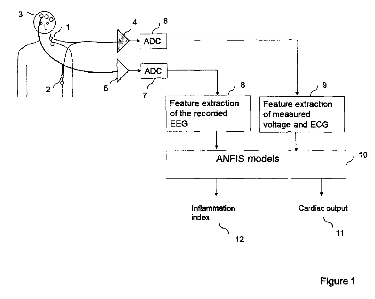
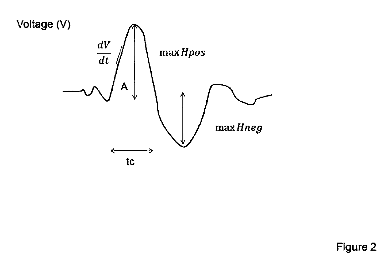
[0040]The novelty of this patent is the combination of several parameters extracted from the voltage plethysmographic curve and the heart rate variability. The voltage plethysmographic curve is achieved by applying a constant current of 400 uA between the upper and lowest electrodes on the thorax, see FIG. 1, (1) and (2). The voltage plethysmographic curve is also referred to as the voltage plethysmogram (VP) or the voltage curve. The voltage curves are achieved for each heart beat, consecutive curves have normally a similar morphology. The voltage is measured between the electrodes adjacent to upper and lowest electrodes (inner electrodes). The current will seek the path with the lowest impedance, i.e. the blood filled aorta. Hence the more blood present the impedance will be lower and consequently the voltage as well. The voltage curve and the ECG are recorded from the same electrodes (1) (2), amplified (4), digitized (6). Features are extracted from ...


PUM


Abstract

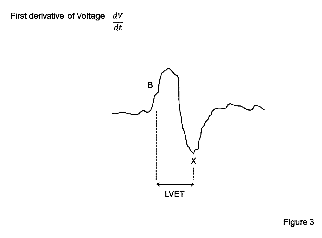
Apparatus for determining stroke volume, cardiac output and systemic inflammation by fuzzy logic combination of features extracted from a voltage measured over the thorax, electrocardiogram and electroencephalogram comprising a) sensor for measuring electroencephalogram; b) sensor for measuring the electrocardiogram; c) calculating the heart rate variability from the electrocardiogram; d) two electrodes (1, 2) for measuring the voltage generated by a constant current generator in two other electrodes (1, 2); e) calculating the area under the curve of voltage for each heart-beat; f) calculating the derivative of the voltage curve; g) calculating the transfer entropy between EEG and ECG; h) combining at least 3 extracted parameters using an Adaptive Neuro Fuzzy inference system (ANFIS) or any other fuzzy reasoner into a final index of cardiac output; i) combining at least 3 extracted parameters using an Adaptive Neuro Fuzzy Inference System or any other fuzzy reasoner into a final index of systemic inflammation.

Description

CROSS-REFERENCE TO RELATED APPLICATIONS[0001]The present application is a U.S. national stage application of International Patent Application No. PCT / DK2014 / 000061, filed Dec. 15, 2014, which claims the benefit of and priority to Danish Patent Application No. PA 2013300389, filed Dec. 13, 2013, both of which are incorporated by reference herein in their entireties.INTRODUCTION[0002]As the patient's haemodynamic status may change rapidly, continuous monitoring of cardiac output will provide information allowing rapid adjustment of therapy. For more than three decades the pulmonary artery catheter (PAC) thermodilution method has been generally accepted and is still the clinical standard to which all other methods are compared.[0003]The reference method for monitoring cardiac output (CO) is the pulmonary artery catheter (PAC). The PAC is also, termed a Swan-Ganz catheter, named after the inventors of the technique. The PAC is introduced into vena cava, and then fed through the heart an...

Claims


Application Information

Patent Timeline
Patent Type & Authority Patents(United States)
IPC IPC(8): A61B5/02G16H50/30G16H50/20G16H40/63A61B5/0476A61B5/04A61B5/00A61B5/0205A61B5/0295A61B5/0245A61B5/024
CPCA61B5/04012A61B5/0205A61B5/0245A61B5/0295G16H50/30A61B5/0476A61B5/7264G16H40/63G16H50/20A61B5/02405A61B5/316A61B5/369
Inventor JENSEN, ERIK WEBER
Owner QUANTIUM MEDICAL SL
Learn More