Automated measurement of brain injury indices using brain CT images, injury data, and machine learning

a brain injury and indices technology, applied in the field of automatic measurement of brain injury indices using brain ct images, injury data, machine learning, etc., to achieve the effect of accurate detection of brain tissue shifting

Active Publication Date: 2019-05-28
VIRGINIA COMMONWEALTH UNIV
View PDF7 Cites 3 Cited by
  • Summary
  • Abstract
  • Description
  • Claims
  • Application Information

AI Technical Summary

Benefits of technology

[0006]An embodiment of the invention provides novel image processing techniques and computer vision methods, as well as expert clinical knowledge, combined to quantitively mimic how physicians read and interpret CT scans in order to more accurately detect shifting of brain tissue, i.e., “midline shift”, or a significant change in the size of lateral ventricles.
[0008]The method implemented first detects the ideal midline based on anatomical features in the brain, bones, and tissues. The ideal midline is an imaginary midline if there were no injuries. Image processing techniques are designed to detect the protrusion in the anterior part of the bone and the falx cerebri in the posterior region of the brain tissue. These anatomical features are exactly the ones inspected by physicians in visual inspection of CT scans. The difficulty of detecting these anatomical features is that there are many other brain structures in the CT images which may interfere or mislead this process. As such, prior knowledge on anatomical structure is used to address this issue. Specifically, prior knowledge is used to approximately identify the regions of these structures and then refine the search using advanced signal processing method. This approach provides both high speed and high accuracy.
[0010]Once the segmentation process is completed, the actual midline is estimated based on these feature points. Using shape match to estimate the actual midline provides a robust and accurate process, which successfully deals a very wide range of pathological cases. After both ideal midline and actual midline are detected, the shift or deformation can be calculated. This shift indicates the midline deformation around the ventricle system.
[0012]The invention provides accurate detection of ideal midline, the use of both anatomical features and spatial templates derived from MRI images to identify ventricles from pathological CT images, and a novel multiple regions shape matching algorithm which provides a very robust actual midline estimation method to deal with diverse ventricle shapes. The invention provides a practical, accurate, automated method for measurement of midline shift from CT images, accurate detection of ventricles from CT image, accurate and non-invasive estimation of ICP, accurate noninvasive screening / diagnostics as well as treatment planning. The application of the technique according to the invention can help physicians avoid time consuming, invasive, risky, and costly alternatives.

Problems solved by technology

The difficulty of detecting these anatomical features is that there are many other brain structures in the CT images which may interfere or mislead this process.

Method used


Image

  • Automated measurement of brain injury indices using brain CT images, injury data, and machine learning
  • Automated measurement of brain injury indices using brain CT images, injury data, and machine learning
  • Automated measurement of brain injury indices using brain CT images, injury data, and machine learning

Examples


Embodiment Construction

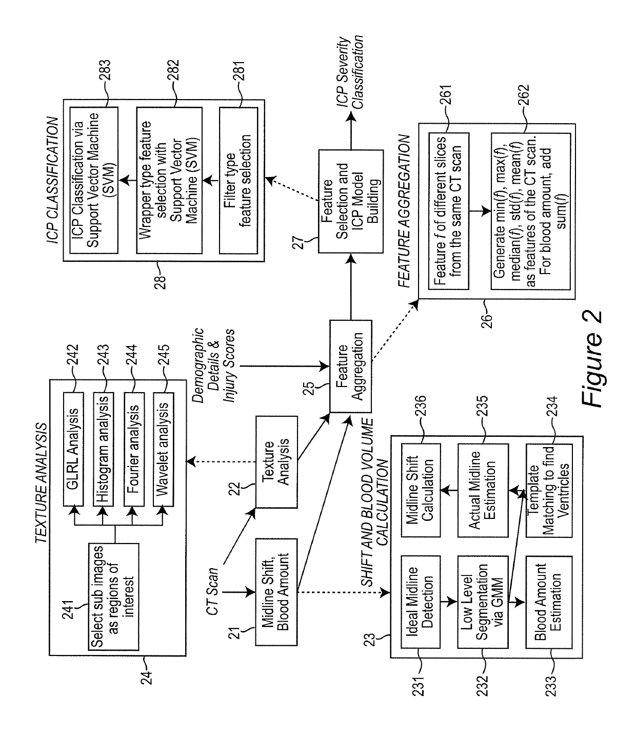
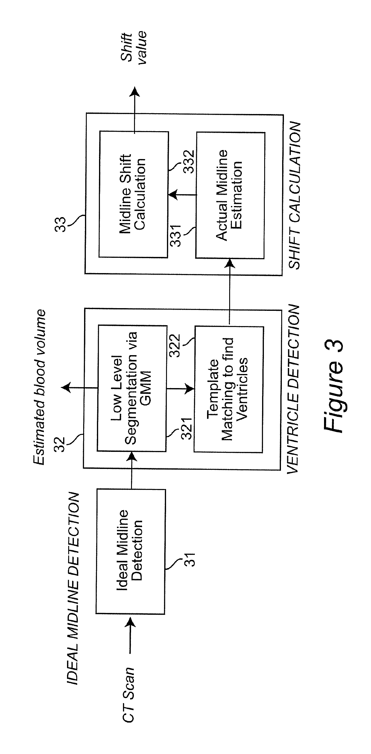
[0029]In clinic settings, the midline shift is widely accepted as an indicator of elevated intracranial pressure (ICP). However, the relation applies only when the midline shift is obvious enough which indicates very high ICP and thus is usually too late for timely treatment. An aim of the present invention is to reveal the relation of ICP with all levels of midline shift as well as other information, such as brain tissue texture in CT images, hematoma volume and pertinent demographic and injury data. The final result is an accurate prediction of ICP to assist medical decisions.

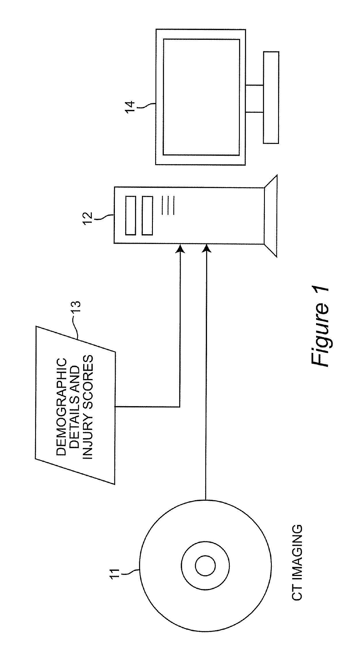
[0030]An embodiment of the invention is described in terms of a decision-support system on which the methods of the invention may be implemented. The system is composed of various signal processing components, imaging processing components, databases and computational interfaces that one of ordinary skill in the computational and signal processing arts will be familiar with. The methods of the invention are d...


PUM


Abstract

A decision-support system and computer implemented method automatically measures the midline shift in a patient's brain using Computed Tomography (CT) images. The decision-support system and computer implemented method applies machine learning methods to features extracted from multiple sources, including midline shift, blood amount, texture pattern and other injury data, to provide a physician an estimate of intracranial pressure (ICP) levels. A hierarchical segmentation method, based on Gaussian Mixture Model (GMM), is used. In this approach, first an Magnetic Resonance Image (MRI) ventricle template, as prior knowledge, is used to estimate the region for each ventricle. Then, by matching the ventricle shape in CT images to the MRI ventricle template set, the corresponding MRI slice is selected. From the shape matching result, the feature points for midline estimation in CT slices, such as the center edge points of the lateral ventricles, are detected. The amount of shift, along with other information such as brain tissue texture features, volume of blood accumulated in the brain, patient demographics, injury information, and features extracted from physiological signals, are used to train a machine learning method to predict a variety of important clinical factors, such as intracranial pressure (ICP), likelihood of success a particular treatment, and the need and / or dosage of particular drugs.

Description

CROSS-REFERENCE TO RELATED APPLICATIONS[0001]This application is a U.S. National Phase of International Application No. PCT / US10 / 27584, filed Mar. 17, 2010, which claims the priority benefit under 35 U.S.C. § 119(e) of U.S. Provisional Application No. 61 / 167,284, filed Apr. 7, 2009, the disclosures of all of which are incorporated herein by reference in their entirety.BACKGROUND OF THE INVENTIONField of the Invention[0002]The present invention generally relates to a decision-support system and computer implemented method to automatically measure the midline shift using Computed Tomography (CT) images and, more particularly, to a decision-support system and computer implemented method that applies machine learning methods to features extracted from multiple sources, including midline shift, blood amount, texture pattern and other injury data, to provide a physician an estimate of intracranial pressure (ICP) levels.Background Description[0003]It is estimated that every year, 1.5 milli...

Claims


Application Information

Patent Timeline
Patent Type & Authority Patents(United States)
IPC IPC(8): A61B5/00G06T7/42G06T7/00G06K9/62G06T7/68G16H50/20G16H30/40G16H50/30
CPCG06K9/629G06T7/0012G06T7/42G06T7/68G16H30/20Y02A90/26G06T2207/10081G06T2207/20056G06T2207/20064G06T2207/30016G16H50/20G06K2209/055Y02A90/10G16H30/40G16H50/30G06V2201/033G06V10/806G06F18/253
Inventor NAJARIAN, KAYVANCHEN, WENANWARD, KEVIN R.
Owner VIRGINIA COMMONWEALTH UNIV
Why Eureka
Try Eureka