Humanized anti-complement factor C1Q antibodies
a technology of complement factor and human antibody, applied in the field of anti-complement factor c1q antibodies, can solve the problems of neurodegeneration and cognitive declin
- Summary
- Abstract
- Description
- Claims
- Application Information
AI Technical Summary
Benefits of technology
Problems solved by technology
Method used
Image
Examples
example 1
n of Humanized Anti-C1q Antibodies
Introduction
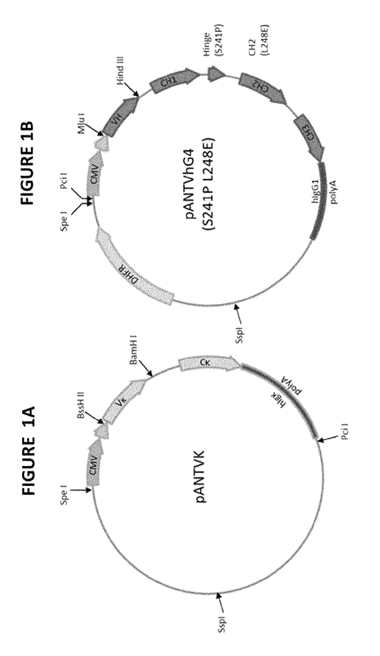
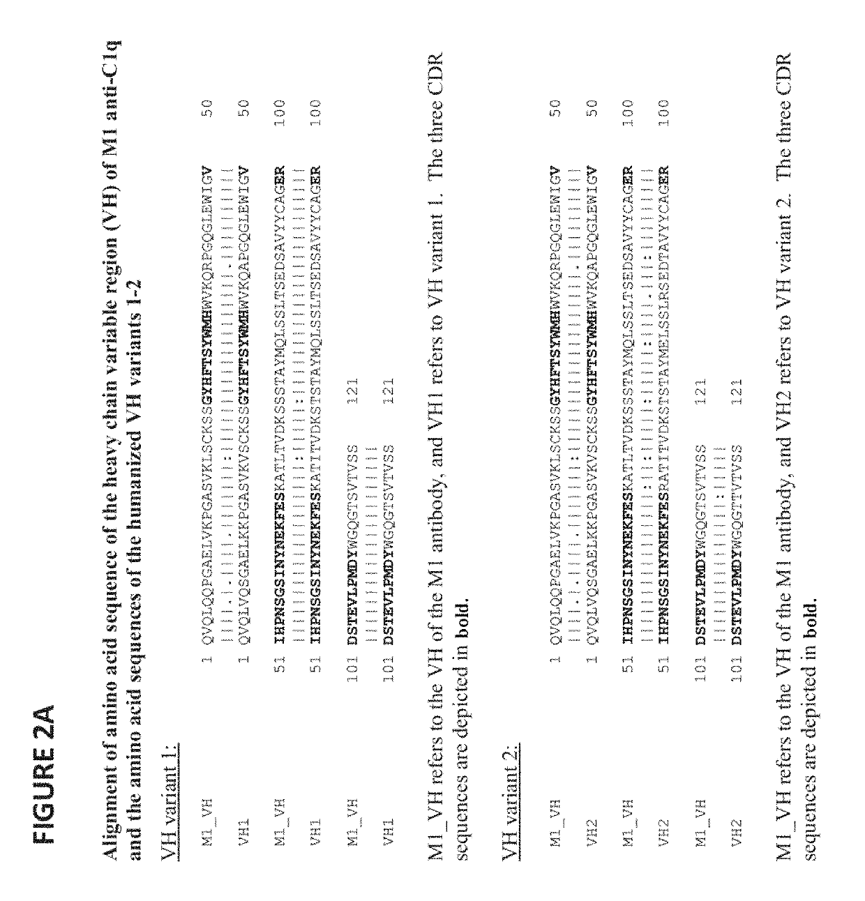
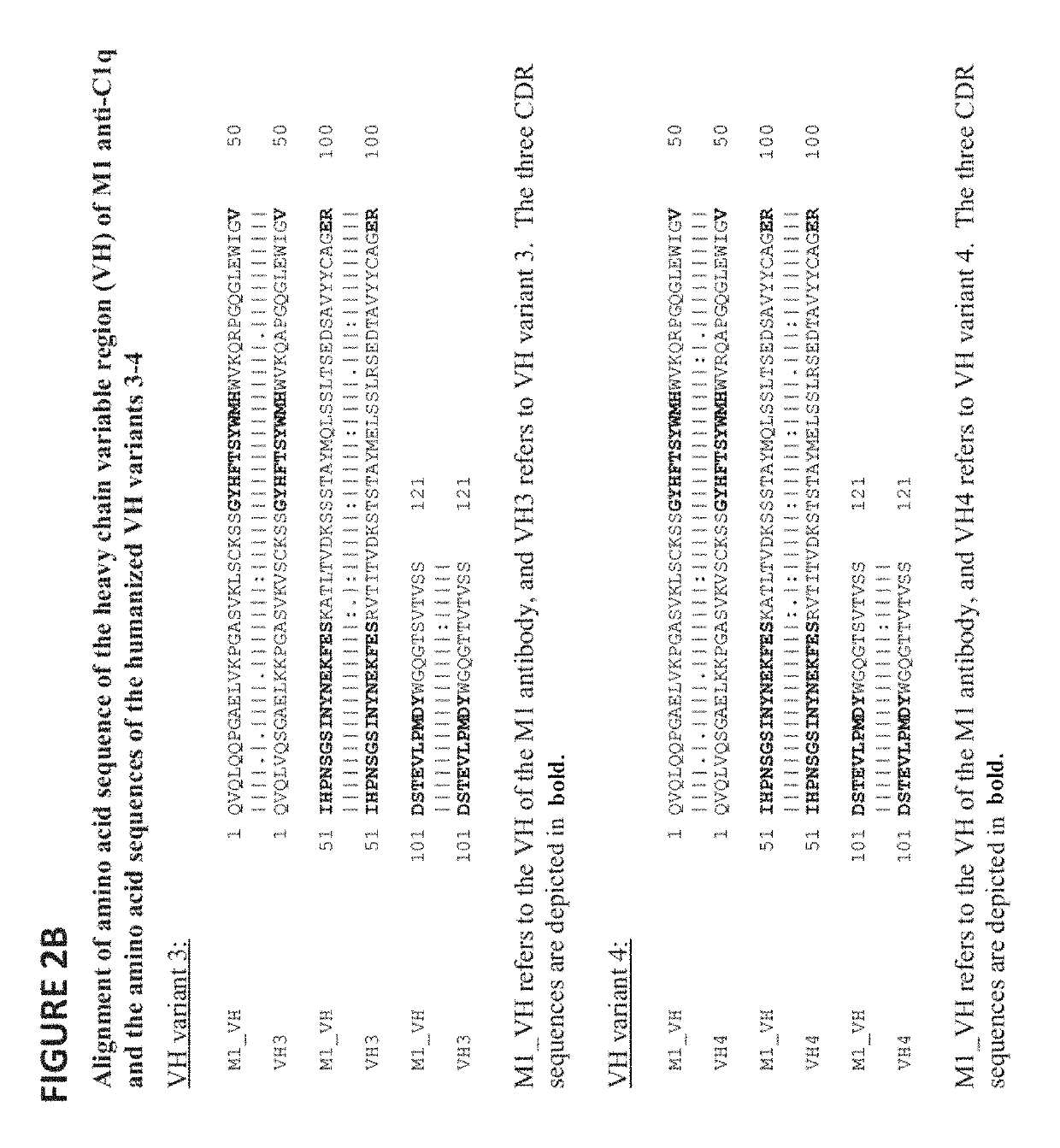
[0261]This example describes the generation of fully humanized antibodies from the murine hybridoma M1 (expressing the mouse anti-human C1q antibody M1). Composite human antibody variable region genes were generated using synthetic oligonucleotides encoding combinations of selected human sequence segments. These were then cloned into vectors encoding human IgG4 (S241P L248E) heavy chain and human kappa light chain. Humanized antibodies were stably expressed in NS0 (mouse myeloma cell-line) cells, Protein A purified and tested for binding to human C1q using a competition ELISA assay against biotinylated murine M1 antibody. Selected antibodies were also tested for binding to mouse C1q using competition ELISA assay against biotinylated chimeric antibody.
Results
Sequencing of Anti-Human C1q V Regions
[0262]RNA was extracted from the hybridoma cell pellet expressing M1 antibody using an RNAqueousR-4PCR kit (Ambion cat. no. AM1914). Initially, R...
example 2
haracterization of Humanized Anti-C1q Antibody VH3 / Vκ3
Introduction
[0345]Immunological biosensors, for example Biacore™ surface plasmon resonance (SPR) instruments, that measure the binding and dissociation of antigen-antibody complexes in real time allow the elucidation of binding kinetics. The rate of dissociation and its subsequent optimization is especially important for biopharmaceutical antibody development.
[0346]The Biacore uses SPR to monitor the interaction between a surface bound molecule ‘ligand’ and its binding partner in solution ‘analyte’, in real time. SPR is an electron charge-density wave phenomenon, which arises at the surface of a metallic layer when light is reflected at the layer under conditions of total internal reflectance. The surface plasmons that are generated are sensitive to any changes in the refractive index of the medium on the opposite side of the metallic layer from the reflected light. Protein-protein interactions occurring at the surface affect the...
example 3
Anti-C1q Antibodies Inhibit Complement-Mediated Hemolysis
[0412]Humanized anti-C1q antibodies were tested in human and rodent hemolytic assays (CH50) for their ability to neutralize C1q and block its activation of the downstream complement cascade.
[0413]The humanized anti-C1q antibodies utilized in this example were produced as described in Example 1. The following humanized antibodies from Example 1 were utilized: the antibody VH1 / Vκ1 (2B12), the antibody VH3 / Vκ3 (5H7), the antibody VH3 / Vκ4 (3F1), and the antibody VH4 / Vκ3 (1D3). The mouse monoclonal antibody M1 (ANN-005) and the chimeric M1antibody (3E2) were also utilized as controls.
[0414]CH50 assays were conducted essentially as described in Current Protocols in Immunology (1994) Supplement 9 Unit 13.1. In brief, 5 microliters (μl) of human serum (Cedarlane, Burlington, N.C.) or 0.625 μl of Wistar rat serum was diluted to 50 μl of GVB buffer (Cedarlane, Burlington, N.C.) and added to 50 μl of the humanized antibodies (1 μg) dilut...
PUM
| Property | Measurement | Unit |
|---|---|---|
| dissociation constant | aaaaa | aaaaa |
| dissociation constant | aaaaa | aaaaa |
| dissociation constant | aaaaa | aaaaa |
Abstract
Description
Claims
Application Information
 - R&D
- Intellectual Property
- Life Sciences
- Materials
- Tech Scout
- Unparalleled Data Quality
- Higher Quality Content
- 60% Fewer Hallucinations
Browse by: Latest US Patents, China's latest patents, Technical Efficacy Thesaurus, Application Domain, Technology Topic, Popular Technical Reports.
© 2025 PatSnap. All rights reserved.Legal|Privacy policy|Modern Slavery Act Transparency Statement|Sitemap|About US| Contact US: help@patsnap.com


