Method and apparatus for cervical image analysis with improved reliability

a technology of image analysis and reliability, applied in the field of methods and apparatus for cervical image analysis with improved reliability, can solve the problems of reducing the transparency of epithelium, reducing the accuracy of false alarms for aceto-white regions, and reducing the endothelial function

Active Publication Date: 2019-12-03
KONINKLJIJKE PHILIPS NV
View PDF7 Cites 0 Cited by
  • Summary
  • Abstract
  • Description
  • Claims
  • Application Information

AI Technical Summary

Benefits of technology

[0009]It is an object of the present invention to provide an improved automated image analysis, by means of which false alarms for aceto-white regions can be reduced.
[0011]According to a first option, transformation zones registered in pre acetic acid and post acetic acid images may be used, e.g. by the zone identifier of claim 1, to identify the region with more than a predetermined minimum change in whiteness due to application of acetic acid. This measure helps in propagating anatomical segmentation of different anatomical regions across different stains.
[0012]According to a second option which can be combined with the first option, color values of an initial color space (e.g. RGB color space) of the transformation zone in the post acetic acid images may be converted, e.g. by the aceto-white identifier of claim 1, to a color space with a color component (e.g., the L component of the Lab color space) that substantially matches human perception of lightness or whiteness. This ensures that proper discrimination in accordance with human perception can be achieved.
[0013]According to a third option which can be combined with the first or second option, pixels in the identified transformation zone may be clustered, e.g. by the aceto-white identifier of claim 1, based on their opacity change and pixels with an opacity change below a predetermined threshold may be removed. Thereby, a straight forward discrimination among dominant and minor opacity changes can be achieved.
[0014]According to a fourth option which can be combined with any one of the above first to third options, clustering based multilevel histogram thresholding may be applied inside the identified transformation zone, e.g. by the aceto-white identifier of claim 1. This measure helps in finding the desired result iteratively

Problems solved by technology

It is also accompanied by a loss of endothelial function, and in some instances considered undesirable.
Acetic acid causes cellular dehydration and reversible coagulation of intracellular proteins, thus reducing the transparency of the epithelium.
This results in temporary whiteness of the epithelium, i.e., aceto-white epithelium.
Unfortunately, not all the areas of aceto-white epithelium indicate the presence of premalignant disease, for example areas of metaplastic epithelium are also aceto-white.
This causes undesirable false alarms for aceto-white regions in automated colposcopic image analysis systems.

Method used


Image

  • Method and apparatus for cervical image analysis with improved reliability
  • Method and apparatus for cervical image analysis with improved reliability
  • Method and apparatus for cervical image analysis with improved reliability

Examples


first embodiment

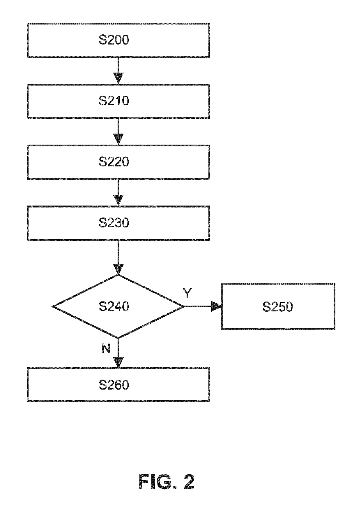
[0035]FIG. 2 shows a schematic flow diagram of an automated image analysis procedure according to a

[0036]In a first step S200, in order to identify the TZ in the cervix automatically and accurately, a post or pre acetic acid image (where the inner most boarder of the TZ i.e. a new SCJ (Sqamo-columnar Junction) is prominent) is registered with Lugol's iodine image (where outer most border of TZ i.e. an old SCJ is prominent) of the same patient at the same level of magnification using e.g. phase congruency and consistent elastic registration. Multi-stain images (obtained by applying saline, acetic acid and Lugol's iodine) having similar magnification levels are registered, which helps in propagating the anatomical segmentation of different anatomical regions across the stains. More specifically, the TZ (i.e. the region between new SCJ and old SCJ) can be identified by registering at least one Lugol's iodine image and one post acetic acid image (i.e. image after application of acetic a...

second embodiment

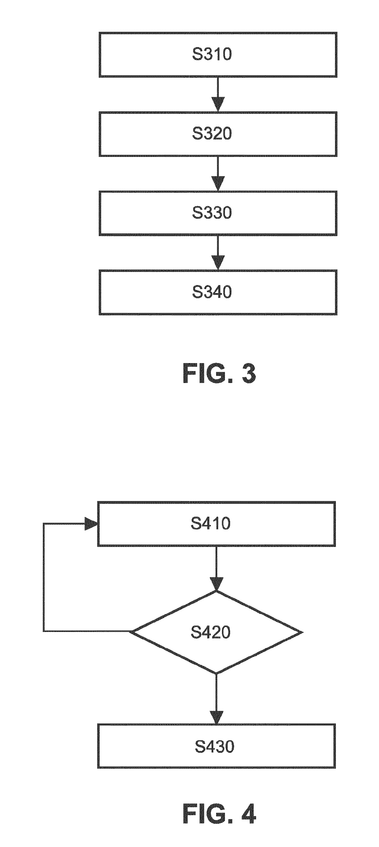
[0044]FIG. 3 shows a schematic flow diagram of a clustering based aceto-white region segmentation procedure according to the

[0045]In step S310, RGB (red, green and blue color) values of the transformation zone in post acetic acid images are converted to the Lab colour space. The Lab color space is a color-opponent space with dimension “L” for lightness and “a” and “b” for the color-opponent dimensions, based on nonlinearly compressed XYZ color space coordinates of the International Commission on Illumination (CIE). The “L” component closely matches human perception of lightness / whiteness. Then, in step S320, pixels are clustered in the transformation zone to two levels of whitish regions, i.e., dominant opacity change and minor opacity change using K-means clustering used to match opaque white and translucent white. K-means clustering is a method of vector quantization and aims at partitioning n observations into k clusters in which each observation belongs to the cluster with the n...

third embodiment

[0046]FIG. 4 shows a schematic flow diagram of an information gain based aceto-white region segmentation procedure as an alternative approach for aceto-whiteness detection according to a

[0047]In step S410, clustering based multilevel histogram thresholding is applied, where a multilevel thresholding process segments grey scale image (inside the TZ region) into several different similar (based on grey scale) regions. The maximum and minimum thresholds are derived from the dataset. Further details are described for example in Seth, S., Naik, S., Jayavanth, S., and Keswarpu, P., “Nucleus Segmentation in Pap-smear Images”, ICBME, 2011. This will help in finding the desired result iteratively. However farther steps is needed to terminate the region grow (with the change of threshold) to achieve the aceto-white regions.

[0048]Then, in step S420, information gain based aceto-white region segmentation is applied. This information gain based homogeneous region identification serves to isolate...


PUM


Abstract

The invention relates to a method and an apparatus for cervical image analysis, wherein a transformation zone is identified in an acetic acid image by registering with its Lugol's iodine counterpart. Then, regions in the transformation zone which show significant changes in whiteness are identified as aceto-white regions and registered with the corresponding Lugol's iodine image, and it is determined if the identified regions in the Lugol's iodine image are iodine negative or positive. Based thereon, the aceto-white region can be categorized as one of metaplasia, inflammation or premalignant lesion region.

Description

CROSS-REFERENCE TO PRIOR APPLICATIONS[0001]This application is the U.S. National Phase application under 35 U.S.C. § 371 of International Application No. PCT / EP2014 / 078181, filed on Dec. 17, 2014, which claims the benefit of European Patent Application No. 13199747.0, filed on Dec. 30, 2013. These applications are hereby incorporated by reference herein.FIELD OF THE INVENTION[0002]The invention relates to the field of apparatuses and methods for semi-automated or automated aceto-whiteness region detection, representation and quantification for any automated cervical image analysis systems.BACKGROUND OF THE INVENTION[0003]When cells are faced with physiological or pathological stresses, they respond by adapting in any of several ways, one of which is metaplasia. It is a benign (i.e. non-cancerous) change that occurs as a response to change of milieu (physiological metaplasia) or chronic physical or chemical irritation (pathological metaplasia). One example of pathological irritation ...

Claims


Application Information

Patent Timeline
Patent Type & Authority Patents(United States)
IPC IPC(8): G06T7/00A61M5/00A61B5/00
CPCA61M5/007G06T7/0012A61B5/4331A61B2576/00G06T2207/30096A61B5/0059G06T2207/10016G06T2207/10024G16H30/40A61B1/303G06T7/136G06T7/174G06T7/194
Inventor SETH, SUBHENDUVAJINEPALLI, PALLAVIKESWARPU, PAYAL
Owner KONINKLJIJKE PHILIPS NV
Why Eureka
Try Eureka