Image-based automated measurement model to predict pelvic organ prolapse

an image-based, automatic technology, applied in the field of pelvic organ prolapse, can solve problems such as difficult identification of particular regions and structures, and achieve the effect of reducing noise and reducing nois

Active Publication Date: 2020-03-17
UNIV OF SOUTH FLORIDA
View PDF2 Cites 3 Cited by
  • Summary
  • Abstract
  • Description
  • Claims
  • Application Information

AI Technical Summary

Benefits of technology

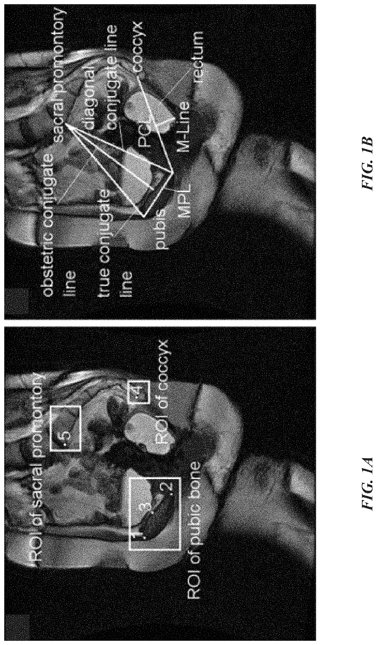
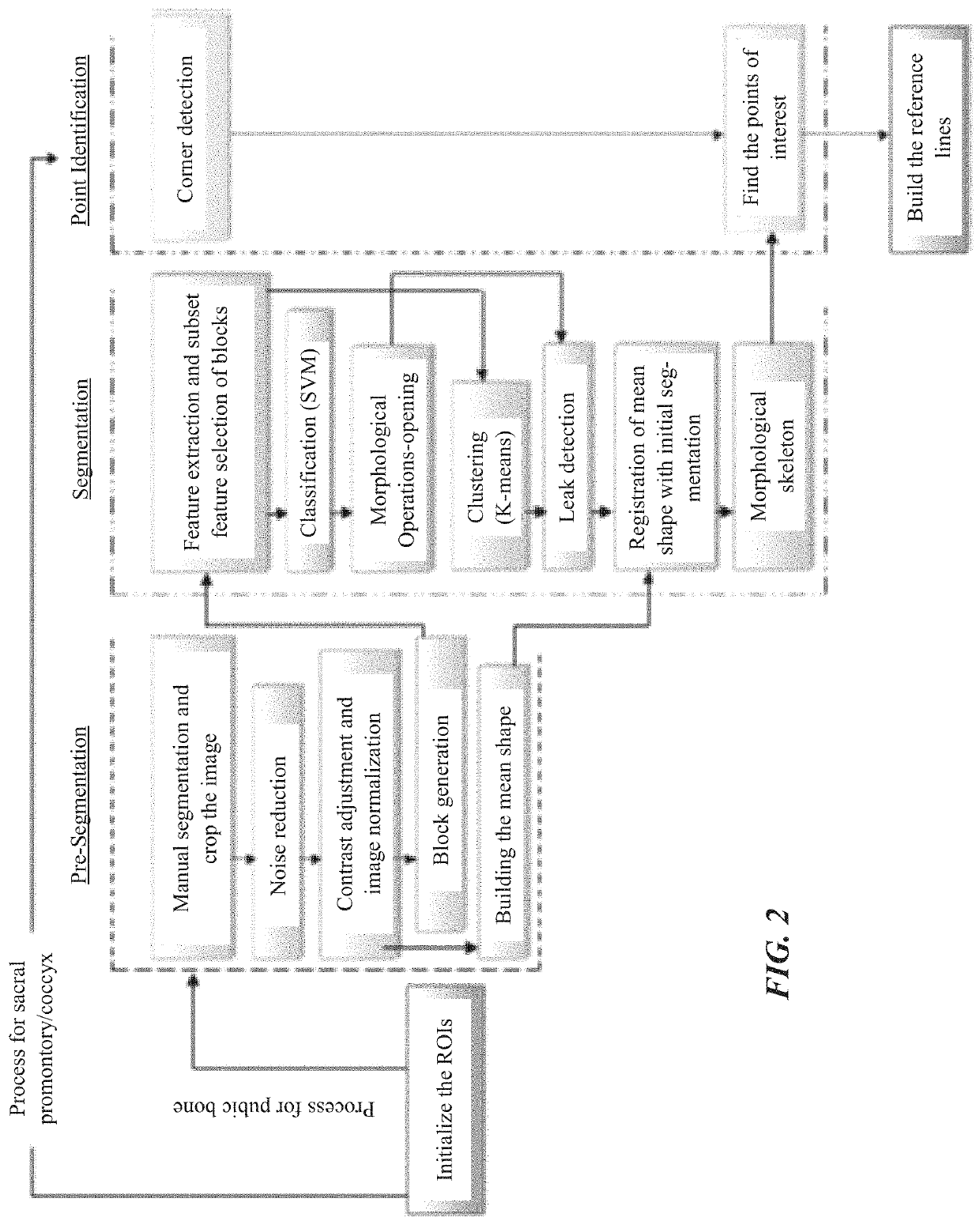
[0031]Identifying the bladder and rectum can be done using a bisecting k-means clustering and morphological opening operations to identify pelvic organs, including the bladder and rectum. The value of k the k-means cluster may be four (4), representing the bone, cartilage, soft tissue and organ, and background regions. In this case, leak detection can be performed to remove clustered cartilage during classification of the bounding boxes. The k-means clustering can also identify the cartilage region and the texture-based classification can identify the bone region, wherein the cartilage and bone regions are combined to find reference points for assessing POP. Alternatively or in addition, identifying the bladder and rectum can include reducing noise and adjusting contrast in the MRI image to normalize the MRI image. In this case, noise is reduced by applying a convolution operation onto the MRI image with smoothing kernel.
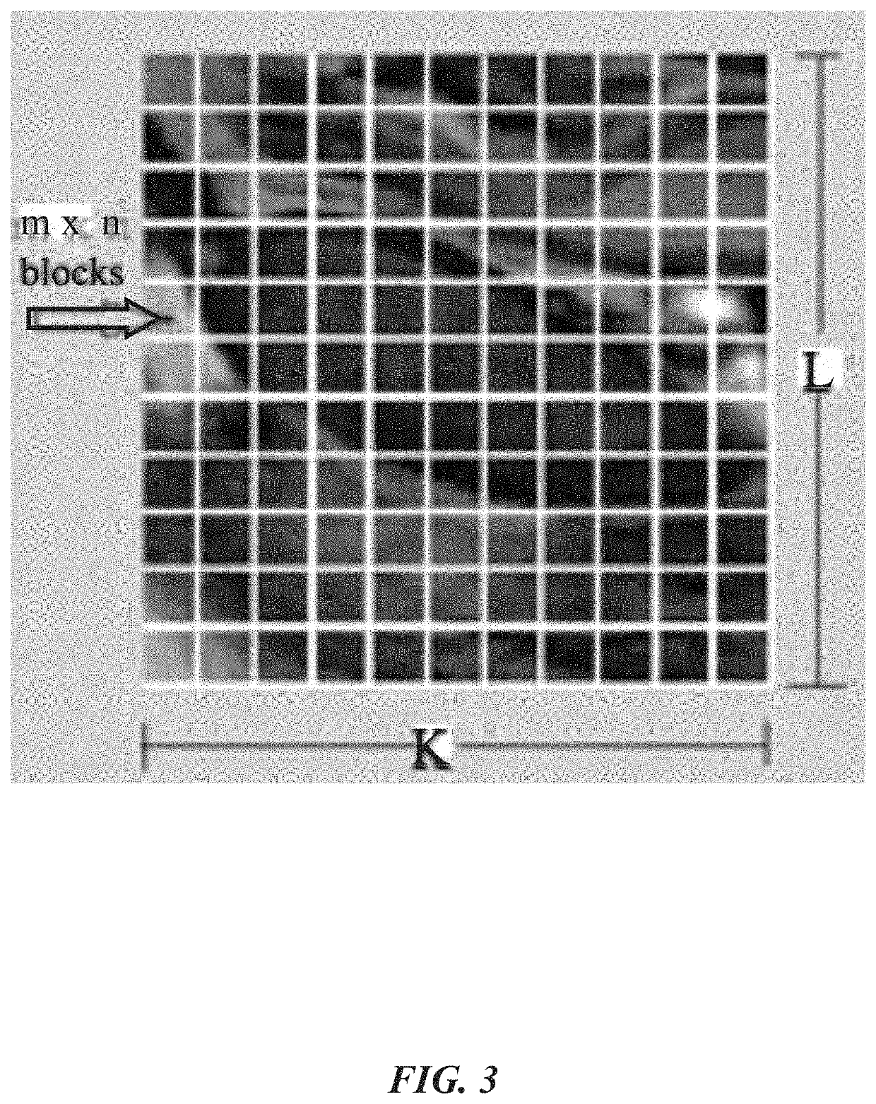
[0032]The keypoints on the pelvic floor may be corner points on said MRI image, such that said corner points can be extracted using a corner point detector. The bounding boxes may be centered at the keypoints, and a size of each bounding box can be based on a mean shape of the pubic bone as determined by the clinical information. If a keypoint is positioned near a boundary of a corresponding bounding box, which thus does not completely enclose an underlying pubic bone structure, the bounding boxes that do completely enclose underlying pubic bone structures are identified by analyzing each bounding box based on Haar-like and texture features. Between identifying and extracting keypoints, any keypoints outside of a vicinity of the bladder and the rectum may be eliminated.
[0036]It is an object of the instant invention provides a quantitative prediction model to better detect POP while increasing understanding of the relationships between MRI-based features and clinical information. It is another object of the instant invention to set the foundation towards a computer-aided decision support system to enable the fusion of image, clinical, and patient data in order to improve the diagnosis of POP and prevent incorrect surgeries through personalized and evidence-based assessment. This will have an enormous impact on pelvic disorder diagnosis, surgical planning, and treatment. The presented system can also be applied to the automated localization and extraction of MRI features for the diagnosis of other diseases where clinical examination is not adequate.

Problems solved by technology

In either case, the method includes providing, inputting, obtaining, or otherwise receiving a patient's pelvic MRI image that may have a low level contrast intensity (and as such difficult to identify particular regions and structures therein).

Method used


Image

  • Image-based automated measurement model to predict pelvic organ prolapse
  • Image-based automated measurement model to predict pelvic organ prolapse
  • Image-based automated measurement model to predict pelvic organ prolapse

Examples


example 1

[0128]The objective of this study is to design, test and validate a prediction model using SVMs and new MRI-based features to differentiate patients with and without POP.

[0129]The main objective of this study is to build a prediction model using SVMs that analyze clinical and new MRI-based features to improve the diagnosis of POP. The current MRI-based features were extracted using the current inventors' previously developed automated pelvic floor measurement model [48, 55]. The significant features, both clinical and MRI-based, were selected using correlation analysis with 95% significance level. The presented prediction model will allow the use of imaging technology to predict the development of POP in predisposed patients, and possibly lead to the development of preventive strategies. Additionally, it is expected that this quantitative prediction model will contribute to a more accurate diagnosis of POP.

[0130]I. Materials and Methods

[0131]This retrospective study used data from 2...

example 2

[0170]A fully automated localization system and methodology is presented herein for automatically locating the bounding boxes of multiple pelvic bone structures on MRI using SVM-based classification and non-linear regression model with global and local information. The model identifies the location of the pelvic bone structures by establishing the association between their relative locations and using local information such as texture features. Results show that the current methodology is able to locate the bone structures of interest accurately.

[0171]More specifically, the methodology described herein first identifies pelvic organs using k-means clustering and morphological opening operations. Then, it uses the spatial relationship between the organs and bone structures to estimate the locations of the structures. The pubic bone is located using the relative location between bones and organs, and texture information. Then, a non-linear regression model is used to predict the locati...


PUM


Abstract

A system and methodology for the automated localization, extraction, and analysis of MRI-based features with clinical information to improve the diagnosis of pelvic organ prolapse (POP). The system can automatically identify reference points for pelvic floor measurements on MRI rapidly and consistent. It provides a prediction model that analyzes the correlation between current and new MRI-based features with clinical information to differentiate patients with and without POP. This system will enable the high throughput analysis of MR images for their correlation with clinical information to better detect POP. The presented system can also be applied to the automated localization and extraction of MRI features for the diagnosis of other diseases where clinical examination is not adequate.

Description

CROSS-REFERENCE TO RELATED APPLICATIONS[0001]This nonprovisional application claims priority to U.S. Provisional Application Ser. No. 62 / 134,702, entitled “Image-Based Automated Measurement Model to Predict Pelvic Organ Prolapse”, filed Mar. 18, 2015 by the same inventors, the entirety of which is incorporated herein by reference.BACKGROUND OF THE INVENTION[0002]1. Field of the Invention[0003]This invention relates, generally, to pelvic organ prolapse. More specifically, it relates to a methodology for analyzing medical images to facilitate prediction and / or diagnosis of pelvic organ prolapse.[0004]2. Brief Description of the Prior Art[0005]Pelvic organ prolapse (POP) is a serious health condition that affects about 30-50% of women in the U.S. [1] and has direct costs of approximately $1 billion / year. POP symptoms are generally nonspecific, and it occurs when a pelvic organ such as bladder, uterus, small bowel and rectum drops from its normal position and pushes against the vaginal ...

Claims


Application Information

Patent Timeline
Patent Type & Authority Patents(United States)
IPC IPC(8): G06T7/00G06K9/46G06K9/62A61B5/055G06V10/50G06V10/764G06V10/774
CPCG06K9/6269G06T7/0012G06K9/4642G06K9/6256A61B5/055G06T2207/30004G06T2207/10088G06K2209/051G06V10/50G06V2201/031G06V10/54G06V10/764G06V10/774G06F18/214G06F18/2411
Inventor ONAL, SINANLAI-YUEN, SUSANA KARINABAO, PAULWEITZENFELD, ALFREDOHART, STUART RICHARD
Owner UNIV OF SOUTH FLORIDA
Why Eureka
Try Eureka