Supercharge Your Innovation With Domain-Expert AI Agents!

Biosensor based on Gβγ-interacting proteins to monitor G-protein activation

a biosensor and gprotein technology, applied in the direction of peptides, enzymology, fluorescence/phosphorescence, etc., can solve the problems of inability to study all the different gproteins using the same detection partners, and the dearth of information available in the field of biosensors for detecting gprotein activation

Active Publication Date: 2020-12-29
UNIV DE MONTREAL
View PDF7 Cites 0 Cited by
  • Summary
  • Abstract
  • Description
  • Claims
  • Application Information

AI Technical Summary

Benefits of technology

The biosensor system provides a sensitive and quantitative assay for monitoring G-protein activation, facilitating the characterization of G-protein coupling profiles and the identification of ligands with biased signaling properties, and can be used in large-scale screening assays and structure-activity relationship studies.

Problems solved by technology

Furthermore, the known methods are not suitable to study all the different G-proteins using the same detection partners.
Even though several approaches are available to support the development of new therapeutically active molecules targeting G-protein activators, the discovery of novel drugs is often limited by the dearth of information available on the precise mechanism of action of those compounds.

Method used


Image

  • Biosensor based on Gβγ-interacting proteins to monitor G-protein activation
  • Biosensor based on Gβγ-interacting proteins to monitor G-protein activation
  • Biosensor based on Gβγ-interacting proteins to monitor G-protein activation

Examples


example 1

Materials and Methods

[0246]Reagents. Angiotensin II (AngII; [Asp-Arg-Val-Tyr-Ile-His-Pro-Phe], SEQ ID NO: 49), poly-ornithine, poly-D-lysine, isoproterenol, rotigotine, epinephrine, norepinephrine, phenylephrine and Pertussis toxin were from Sigma®. u46619 were from Cayman Chemical® (Ann Arbor, Mich.). [Sar1, Ile8]-AngII (SI) and [Asp1, Val5, Gly8]-AngII (DVG) [Sar1-Val5-D-Phe8] AngII (SVdF) and [Sar1-D-Ala8] AngII were synthesized at the University de Sherbrooke (Canada, QC). UBO-Qic (L-threonine,(3R)-N-acetyl-3-hydroxy-L-leucyl-(aR)-a-hydroxybenzenepropanoyl-2,3-idehydro-N-methylalanyl-L-alanyl-N-methyl-L-alanyl-(3R)-3-[[(2S,3R)-3-hydroxy-4-methyl-1-oxo-2-[(1-oxopropyl)amino]pentyl]oxy]-L-leucyl-N,O-dimethyl-,(7→1)-lactone (9CI)) was obtained from Institute for Pharmaceutical Biology of the University of Bonn (Germany). Dulbecco's modified Eagles medium (DMEM), fetal bovine serum, OPTI-MEM®, and other cell culture reagents were purchased from Invitrogen®. Coelenterazine 400a, Coel...

example 2

Results

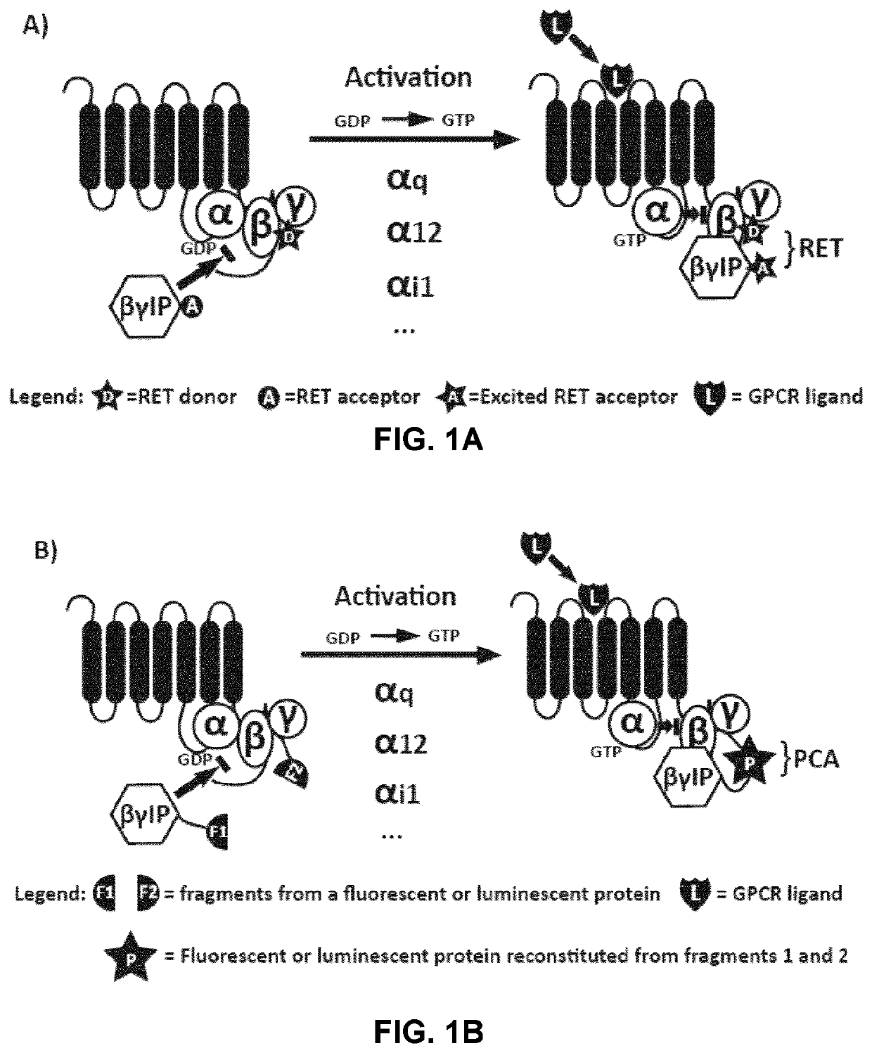
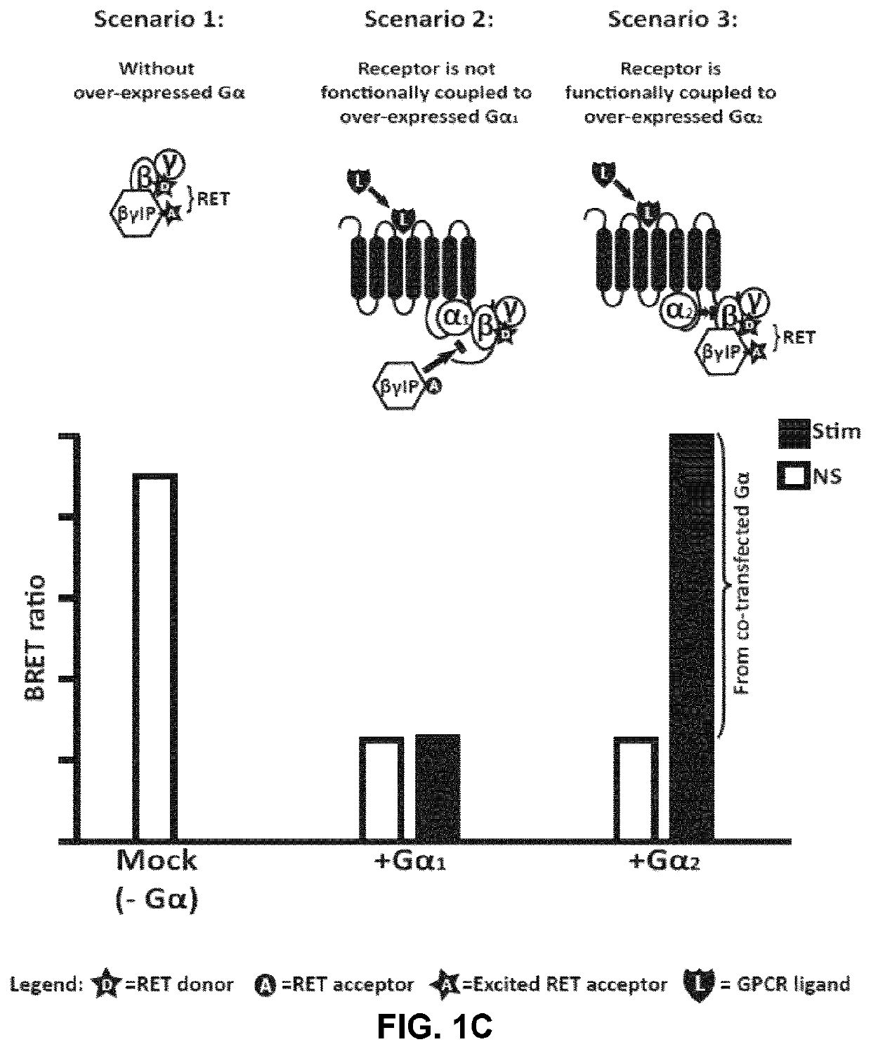
[0260]To study the activation of specific G-proteins by GPCRs, an assay was developed based on the competition between Gα subunits and βγIP for their binding to Gβγ subunits. As depicted in FIGS. 1A and 1B, in the absence of receptor activation, the Gα subunit is tightly bound to the Gβγ dimer, preventing its association with the βγIP. Following receptor stimulation, the GTP-bound Gα dissociates from the Gβγ complex, which is then free to interact with the βγIP. The interaction between the βγIP and the Gβγ, therefore, reflects the G-protein activation. By co-expressing βγIP and Gβγ, each tagged with one of the two components of the detection system, with different subtypes of untagged Gα, it has been possible to determine the coupling profile of a given receptor following its activation. As shows below, different methods of detection may be used to assess the interaction between the βγIP and the Gβγ, such as resonance energy transfer (RET) approaches (FIG. 1A: bioluminescence...


PUM

PropertyMeasurementUnit
wavelengthaaaaaaaaaa
pHaaaaaaaaaa
energy transferaaaaaaaaaa

Abstract

Resonance energy transfer (RET)- or protein-fragment complement assay (PCA)-based biosensors useful for assessing the activity of G-proteins are described. These biosensors are based on the competition between the Gα subunit and a Gβγ interacting protein (βγ IP) for the binding to the Gβγ dimer. These biosensors comprises (1) a βγ IP and (2) a Gβ or Gγ protein; a GPCR; or a plasma membrane targeting domain, fused to suitable RET or PCA tags. Methods using such biosensors for different applications, including the identification of agents that modulates G-protein activity or for the characterization of GPCR signaling / regulation, such as G-protein preferences and activation profiles of GPCRs, are also described.

Description

CROSS REFERENCE TO RELATED APPLICATIONS[0001]This application claims the benefit of U.S. provisional application Ser. No. 62 / 063,622, filed on Oct. 14, 2014, which is incorporated herein by reference in its entirety.TECHNICAL FIELD[0002]The present disclosure relates to the monitoring of G-protein activation, and more specifically to a signaling biosensor for detecting G-protein activation.BACKGROUND ART[0003]Hetero-trimeric G-proteins consisting of three subunits α, β and γ, relays the information provided by G-protein-coupled receptors (GPCRs) to various intracellular effectors. In the absence of stimulation, the α-subunit of the G-protein is in complex with a GDP (guanosine diphosphate) molecule. The conformational change that follows receptor activation by a ligand, promotes the phosphorylation of the GDP molecule into a GTP (guanosine triphosphate). The GTP-bound Gα subunit dissociates from the Gβγ subunits, both of which are then available to interact with downstream effectors...

Claims


Application Information

Patent Timeline
Patent Type & Authority Patents(United States)
IPC IPC(8): C12N9/02G01N21/64G01N33/542C07K14/72C12Q1/66C07K14/47G01N33/566
CPCC12Q1/66C12Y113/12007G01N33/566C07K14/723C07K14/4722G01N33/542G01N21/6428C12N9/0069G01N2333/726C07K2319/01G01N2021/6439G01N2333/4719C07K2319/60G01N2500/02C07K2319/00
Inventor BOUVIER, MICHELLE GOUILL, CHRISTIANHOGUE, MIREILLELUKASHEVA, VIKTORIYA
Owner UNIV DE MONTREAL
Features
  • R&D
  • Intellectual Property
  • Life Sciences
  • Materials
  • Tech Scout
  • Unparalleled Data Quality
  • Higher Quality Content
  • 60% Fewer Hallucinations
Learn More