
Hollow-fiber membrane and hollow-fiber module for cell culture

a cell culture and hollow fiber technology, applied in the field of hollow fiber membranes, can solve the problems of high risk of biological contamination, inability to differentiate into all organs and tissues, and inability to differentiate into specific tissues, and achieve the effect of safe, easy and efficient culturing of various kinds of cells

Active Publication Date: 2021-01-26
TOYOBO CO LTD
View PDF18 Cites 0 Cited by
  • Summary
  • Abstract
  • Description
  • Claims
  • Application Information

AI Technical Summary

Benefits of technology

[0018]According to the present invention, it is possible to provide a hollow-fiber membrane as a culture substrate which does not require a coating treatment with a cell adhesion factor. In addition, when the hollow-fiber membrane of the present invention is used, it is possible to construct and provide a cell culture system capable of safely, easily and efficiently culturing various kinds of cells such as mesenchymal stem cells.

Problems solved by technology

On the other hand, somatic stem cells cannot differentiate into all organs and tissues, and differentiate into specific tissues and organs.
However, there exist an extremely small number of such somatic stem cells in tissues.
However, in cell culture operations that are generally carried out, there is the problem that the risk of biological contamination is high, and costs associated with personnel expenses are high, and therefore it is required to safely and inexpensively culture stem cells for further development of cell transplantation therapy.
Incidentally, cells adhesive in a living body cannot grow or survive outside the living body unless the cells adhere to a culture substrate.
However, when a culture substrate is coated with such a cell adhesion factor, material costs of the adhesion factor and operation costs are increased.
In addition to the problem of cost increase, there arises the additional risk of unknown infection when the cell adhesion factor is an animal-derived component.
In recent years, the risk of infection has been reduced by using a cell adhesion factor produced by gene-recombinant Escherichia coli, but in this production, complicated operations such as tracking of a source of each medium component that is used in a process for culturing recombinant Escherichia coli may be required.

Method used

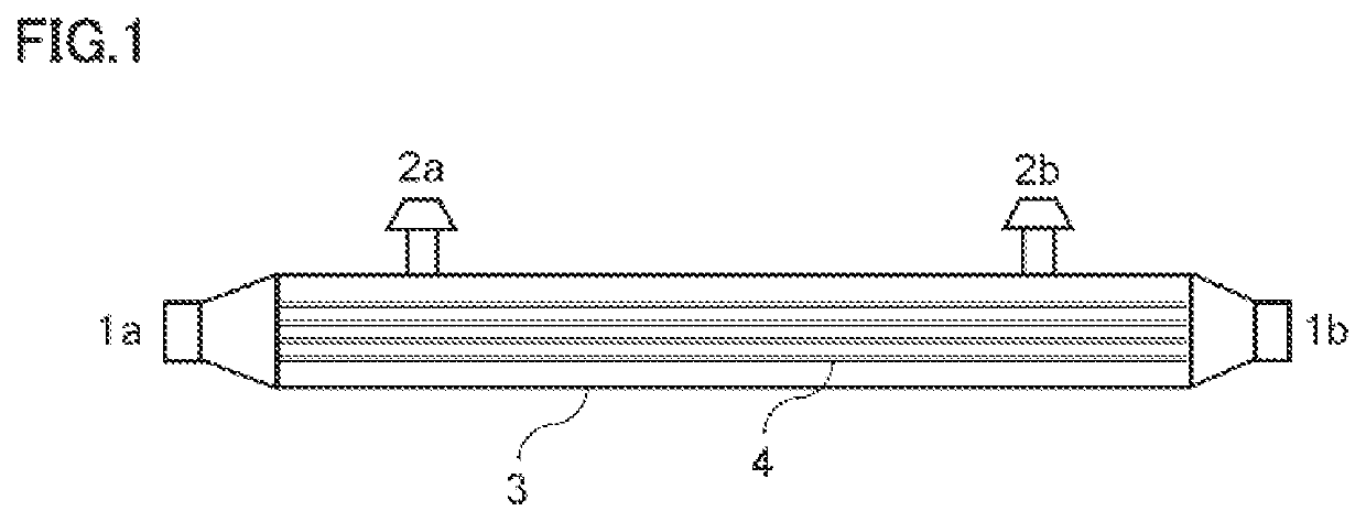
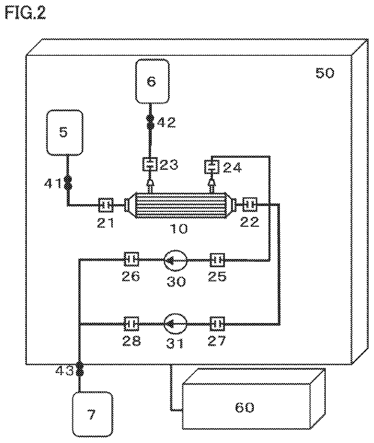

Image

  • Hollow-fiber membrane and hollow-fiber module for cell culture
  • Hollow-fiber membrane and hollow-fiber module for cell culture
  • Hollow-fiber membrane and hollow-fiber module for cell culture

Examples


example 1

(Preparation of Hollow-Fiber Membrane and Hollow-Fiber Module 1)

[0067]20% by mass of polyether sulfone (PES) (4800 P manufactured by Sumitomo Chemical Company, Limited), 0.2% by mass of polyvinyl pyrrolidone (PVP) (K-90 manufactured by BASF Company), 35.91% by mass of N-methyl pyrrolidone (NMP), and 43.89% by mass of triethylene glycol (TEG) were mixed and dissolved, the resulting solution was defoamed to obtain a deposition solution, a mixed liquid of 13.5% by mass of NMP, 16.5% by mass of TEG and 70% by mass of water was provided as a core liquid, the deposition solution and the core liquid were discharged, respectively, from the outside and the inside of a double-tube orifice heated to 70° C., and guided through a 30 cm idle running section into a coagulation bath including water at 75° C., so that a hollow-fiber membrane was formed. The hollow-fiber membrane was washed with water, and then wound up in the form of a bundle. The fiber bundle was cut, and then subjected to forced-a...

example 2

(Preparation of Hollow-Fiber Membrane and Hollow-Fiber Module 2)

[0069]20% by mass of PES, 0.5% by mass of PVP, 35.77% by mass of NMP and 43.73% by mass of TEG were mixed and dissolved, and the resulting solution was defoamed to obtain a deposition solution, a mixed liquid of 13.5% by mass of NMP, 16.5% by mass of TEG and 70% by mass of water was provided as a core liquid, the deposition solution and the core liquid were discharged, respectively, from the outside and the inside of a double-tube orifice heated to 70° C., and guided through a 30 cm idle running section into a coagulation bath including water at 75° C., so that a hollow-fiber membrane was formed. The hollow-fiber membrane was washed with water, and then wound up in the form of a bundle. The fiber bundle was cut, and then subjected to forced-air drying at 60° C. The dried hollow-fiber membrane had an inner diameter of 200 μm, an outer diameter of 300 μm and a thickness of 50 μm.

[0070]Using the obtained hollow-fiber membr...

example 3

(Preparation of Hollow-Fiber Membrane and Hollow-Fiber Module 3)

[0071]20% by mass of PES, 1.0% by mass of PVP, 35.55% by mass of NMP and 43.45% by mass of TEG were mixed and dissolved, the resulting solution was defoamed to obtain a deposition solution, a mixed liquid of 13.5% by mass of NMP, 16.5% by mass of TEG and 70% by mass of water was provided as a core liquid, the deposition solution and the core liquid were discharged, respectively, from the outside and the inside of a double-tube orifice heated to 70° C., and guided through a 30 cm idle running section into a coagulation bath including water at 75° C., so that a hollow-fiber membrane was formed. The hollow-fiber membrane was washed with water, and then wound up in the form of a bundle. The fiber bundle was cut, and then subjected to forced-air drying at 60° C. The obtained hollow-fiber membrane had an inner diameter of 200 μm, an outer diameter of 300 μm and a thickness of 50 μm.

[0072]Next, Using the obtained hollow-fiber ...


PUM

PropertyMeasurementUnit
contact angleaaaaaaaaaa
inner diameteraaaaaaaaaa

Abstract

An object of the present invention is to provide a hollow-fiber membrane which does not require a coating treatment with a cell adhesion factor or surface modification by an electron beam or the like and which is capable of adhering and culturing cells, and a method for culturing cells using the hollow-fiber membrane. A hollow-fiber membrane for cell culture which is to be used as a culture substrate for adhesive cells, in which the hollow-fiber membrane includes a hydrophobic polymer and a hydrophilic polymer, the content of the hydrophilic polymer in the whole hollow-fiber membrane is more than 0% by mass and less than 1% by mass, and the content of the hydrophilic polymer on a surface of the hollow-fiber membrane is more than 0% by mass and less than 10% by mass.

Description

TECHNICAL FIELD[0001]The present invention relates to a hollow-fiber membrane to be used as a culture substrate for culturing cells.BACKGROUND ART[0002]Stem cells are cells that can form organs and tissues, and are considered to exist present in most organs and tissues even in adults. Among stem cells, embryonic stem cells (ES cells) and induced pluripotent stem cells (iPS cells) are pluripotent cells, and are capable of being differentiating into all tissues and organs. On the other hand, somatic stem cells cannot differentiate into all organs and tissues, and differentiate into specific tissues and organs. Somatic stem cells collectable from human tissues are attracting attention as transplanting cells to be used in cell transplantation therapy because they can be collected from patients themselves, and there is no fear of rejection.[0003]As somatic stem cells in humans, mesenchymal stem cells, hematopoietic stem cells, neural stem cells, myocardial stem cells, pancreatic stem cel...

Claims


Application Information

Patent Timeline
Patent Type & Authority Patents(United States)
IPC IPC(8): B01D69/02B01D63/04B01D69/08B01D71/68C12M1/12B01D71/44
CPCB01D69/02B01D63/04B01D69/08B01D71/44B01D71/68C12M25/10C12M25/12B01D2325/36B01D2325/38B01D71/441
Inventor HIRAOKA, MAKIKOYAMAGUCHI, TATSUYAKATO, NORIAKI
Owner TOYOBO CO LTD
Learn More