Medical image processing apparatus, method, and program
a medical image processing and image processing technology, applied in the field of medical image processing apparatus, medical image processing method, medical image processing program, can solve the problems of erroneously detecting a region where subarachnoid hemorrhage cannot occur originally, delay in treatment, etc., and achieve the effect of accurately diagnosing subarachnoid hemorrhage and high accuracy
- Summary
- Abstract
- Description
- Claims
- Application Information
AI Technical Summary
Benefits of technology
Problems solved by technology
Method used
Image
Examples
first embodiment
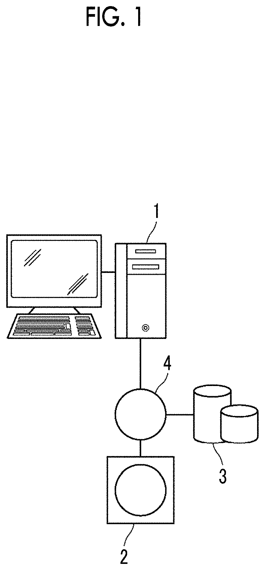
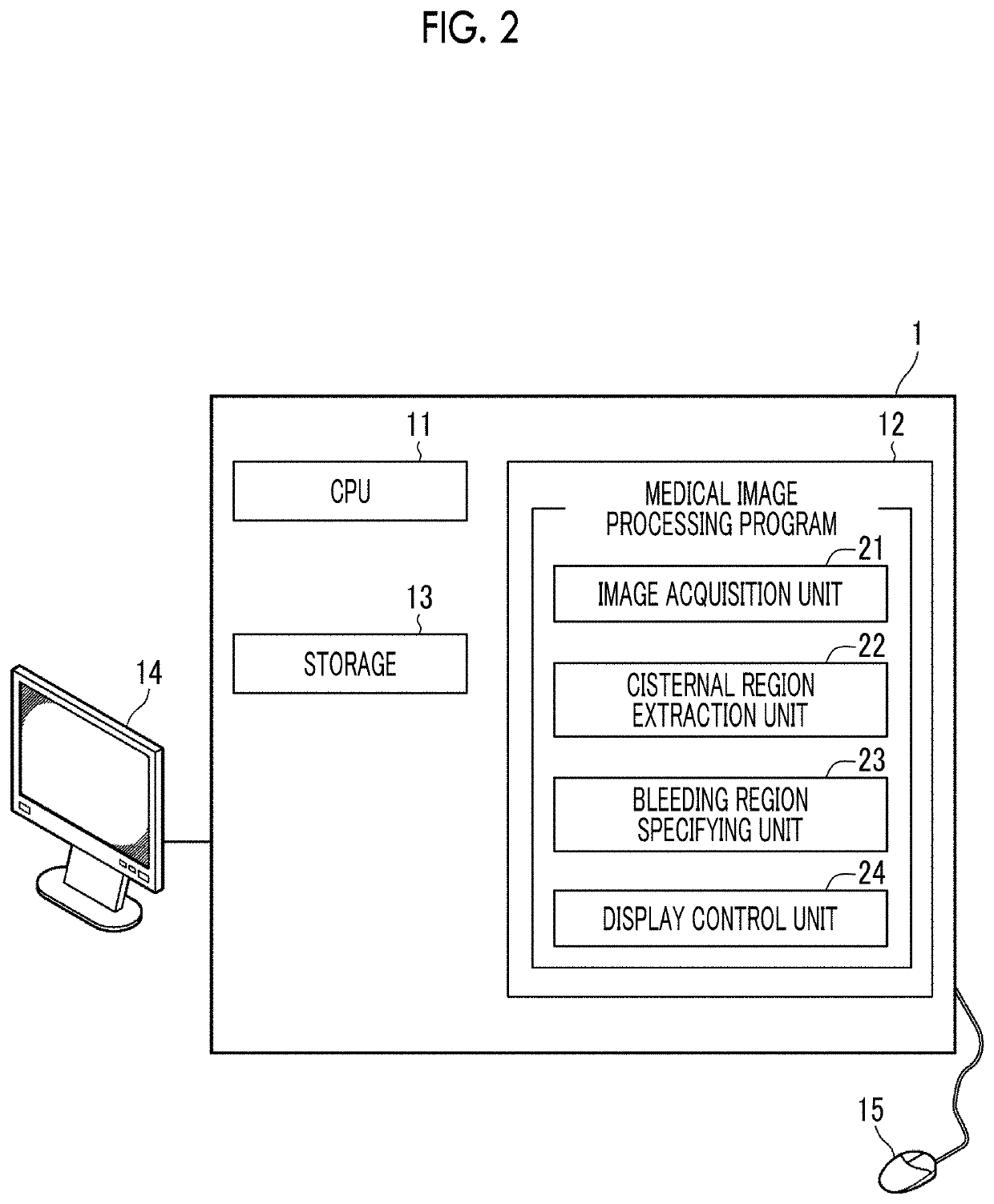
[0034]Hereinafter, embodiments of the invention will be described with reference to the diagrams. FIG. 1 is a hardware configuration diagram showing the outline of a diagnostic support system to which a medical image processing apparatus according to the invention is applied. As shown in FIG. 1, in the diagnostic support system, a medical image processing apparatus 1 according to the present embodiment, a three-dimensional image capturing apparatus 2, and an image storage server 3 are communicably connected to each other through a network 4.
[0035]The three-dimensional image capturing apparatus 2 is an apparatus that generates a three-dimensional image showing a part, which is an examination target part of a subject, as a medical image by imaging the part. Specifically, the three-dimensional image capturing apparatus 2 is a CT apparatus, an MRI apparatus, a positron emission tomography (PET) apparatus, or the like. The medical image generated by the three-dimensional image capturing ...
third embodiment
[0071]In the third embodiment, for machine learning of the discriminator 25, a number of differences between the first signal value distribution and the second signal value distribution of a cisternal region, which is known to be a bleeding region, and a number of differences between the first signal value distribution and the second signal value distribution of a cisternal region, which is not a bleeding region, are prepared. Then, machine learning is performed using the number of differences, thereby generating the discriminator 25. As a method of machine learning, any known method using a logistic circuit, a support vector machine, and the like can be used. By performing learning in this manner, the discriminator 25 outputs a determination result as to whether or not the cisternal region 32 is a bleeding region in a case where the difference between the first signal value distribution of the cisternal region 32 extracted from the brain image B0 and the second signal value distrib...
PUM
 Abstract
Description
Claims
Application Information
 - Generate Ideas
- Intellectual Property
- Life Sciences
- Materials
- Tech Scout
- Unparalleled Data Quality
- Higher Quality Content
- 60% Fewer Hallucinations
Browse by: Latest US Patents, China's latest patents, Technical Efficacy Thesaurus, Application Domain, Technology Topic, Popular Technical Reports.
© 2025 PatSnap. All rights reserved.Legal|Privacy policy|Modern Slavery Act Transparency Statement|Sitemap|About US| Contact US: help@patsnap.com


