
Medical image processing apparatus, method, and program

Active Publication Date: 2019-11-14
FUJIFILM CORP
View PDF0 Cites 0 Cited by
  • Summary
  • Abstract
  • Description
  • Claims
  • Application Information

AI Technical Summary

Benefits of technology

The invention aims to accurately extract a bleeding region suspected of subarachnoid hemorrhage using a brain image, such as a CT image. By extracting a cisternal region from the brain image and specifying a bleeding region based on the distribution of signal values, the invention enables the accurate diagnosis of subarachnoid hemorrhage using the brain image.

Problems solved by technology

Therefore, in a case where a non-specialist in the brain makes a diagnosis, a bleeding region may be overlooked and accordingly, the start of treatment may be delayed or no treatment may be performed.
However, since the entire region of the brain is a target in both the methods, there is a possibility of erroneously detecting a region where subarachnoid hemorrhage cannot occur originally.

Method used


Image

  • Medical image processing apparatus, method, and program
  • Medical image processing apparatus, method, and program
  • Medical image processing apparatus, method, and program

Examples


first embodiment

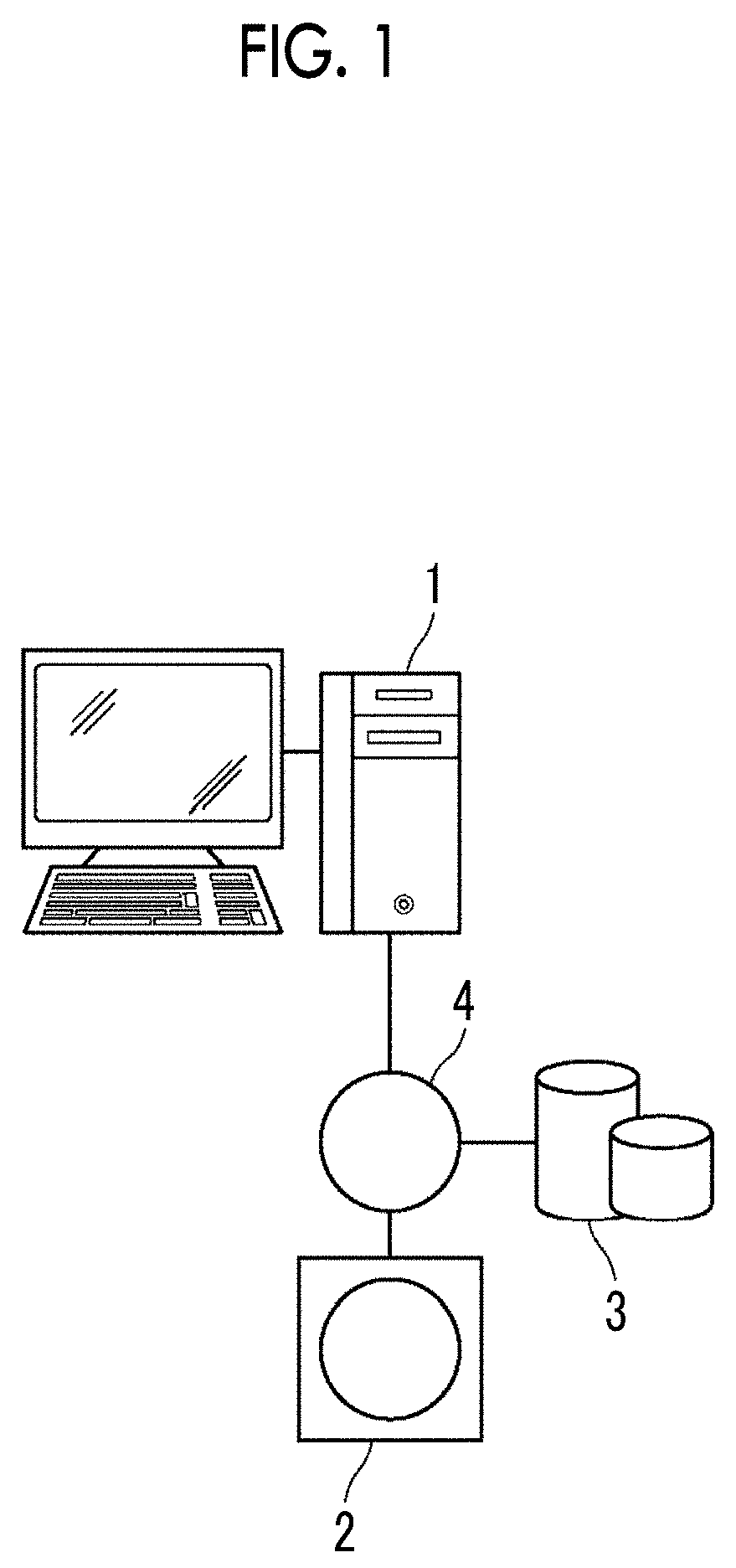
[0034]Hereinafter, embodiments of the invention will be described with reference to the diagrams. FIG. 1 is a hardware configuration diagram showing the outline of a diagnostic support system to which a medical image processing apparatus according to the invention is applied. As shown in FIG. 1, in the diagnostic support system, a medical image processing apparatus 1 according to the present embodiment, a three-dimensional image capturing apparatus 2, and an image storage server 3 are communicably connected to each other through a network 4.

[0035]The three-dimensional image capturing apparatus 2 is an apparatus that generates a three-dimensional image showing a part, which is an examination target part of a subject, as a medical image by imaging the part. Specifically, the three-dimensional image capturing apparatus 2 is a CT apparatus, an MRI apparatus, a positron emission tomography (PET) apparatus, or the like. The medical image generated by the three-dimensional image capturing ...

third embodiment

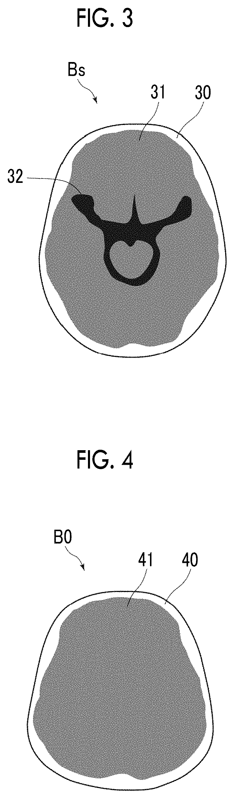
[0071]In the third embodiment, for machine learning of the discriminator 25, a number of differences between the first signal value distribution and the second signal value distribution of a cisternal region, which is known to be a bleeding region, and a number of differences between the first signal value distribution and the second signal value distribution of a cisternal region, which is not a bleeding region, are prepared. Then, machine learning is performed using the number of differences, thereby generating the discriminator 25. As a method of machine learning, any known method using a logistic circuit, a support vector machine, and the like can be used. By performing learning in this manner, the discriminator 25 outputs a determination result as to whether or not the cisternal region 32 is a bleeding region in a case where the difference between the first signal value distribution of the cisternal region 32 extracted from the brain image B0 and the second signal value distrib...


PUM


Abstract

An image acquisition unit acquires a brain image of a subject. A cisternal region extraction unit extracts a cisternal region from the brain image by performing registration between a standard brain image and the brain image. Then, a bleeding region specifying unit specifies a bleeding region based on a first signal value distribution, which is the signal value distribution of the cisternal region extracted from the brain image, and a second signal value distribution, which is the signal value distribution of a cisternal region of the standard brain image.

Description

CROSS REFERENCE TO RELATED APPLICATIONS[0001]The present application claims priority under 35 U.S.C. § 119 to Japanese Patent Application No.2018-090345 filed on May 9, 2018. The above application is hereby expressly incorporated by reference, in its entirety, into the present application.BACKGROUNDTechnical Field[0002]The present invention relates to a medical image processing apparatus, a medical image processing method, and a medical image processing program for extracting a bleeding region suspected of subarachnoid hemorrhage using a brain image, such as a computed tomography (CT) image of the head.Related Art[0003]In recent years, advances in medical apparatuses, such as CT apparatuses and magnetic resonance imaging (MRI) apparatuses have enabled image diagnosis using high-resolution medical images with higher quality. In particular, in a case where a target part is a brain, a region causing a vascular disorder, such as cerebral infarction and cerebral hemorrhage, can be specif...

Claims


Application Information

Patent Timeline
IPC IPC(8): A61B6/00A61B5/055A61B6/03
CPCG06T7/0012G06T2207/30016A61B6/501G06T2207/10081A61B2576/026A61B6/032A61B5/055A61B6/504A61B6/5217A61B6/5294A61B6/5235G16H50/30G06T7/0014
Inventor TAKEI, MIZUKI
Owner FUJIFILM CORP
Features
  • Generate Ideas
  • Intellectual Property
  • Life Sciences
  • Materials
  • Tech Scout
  • Unparalleled Data Quality
  • Higher Quality Content
  • 60% Fewer Hallucinations
Learn More