
Method for determining ischemic status or assessing stroke onset time of a brain region

a brain region and ischemic status technology, applied in the field of magnetic resonance imaging, can solve the problems of increased medical labor, increased cost and risk, and additional errors for patients

Active Publication Date: 2022-01-25
TAIPEI MEDICAL UNIV
View PDF5 Cites 2 Cited by
  • Summary
  • Abstract
  • Description
  • Claims
  • Application Information

AI Technical Summary

Benefits of technology

This approach allows for rapid and cost-effective identification of ischemic status and stroke onset time, enhancing clinical judgment for surgical interventions by eliminating errors in image processing and reducing the need for MRI contrast agents, thus improving patient outcomes within the critical golden hour.

Problems solved by technology

Furthermore, the patient also needs to bear the cost and risk of MRI contrast agent, and the medical labor rises thereby.
However, such image processing may result in additional errors.
Therefore, the conventional clinical tool for identification and differentiation of infarct core and ischemic penumbra is time-consuming and costly.
In addition, there is not yet a clinical tool for assessing the stroke onset time, and an objective index for the golden hour of acute ischemic stroke is still absent.
Thus the clinical judgement of surgical intervention is difficult.

Method used


Image

  • Method for determining ischemic status or assessing stroke onset time of a brain region
  • Method for determining ischemic status or assessing stroke onset time of a brain region
  • Method for determining ischemic status or assessing stroke onset time of a brain region

Examples


examples

Materials and Methods

Animal Preparations

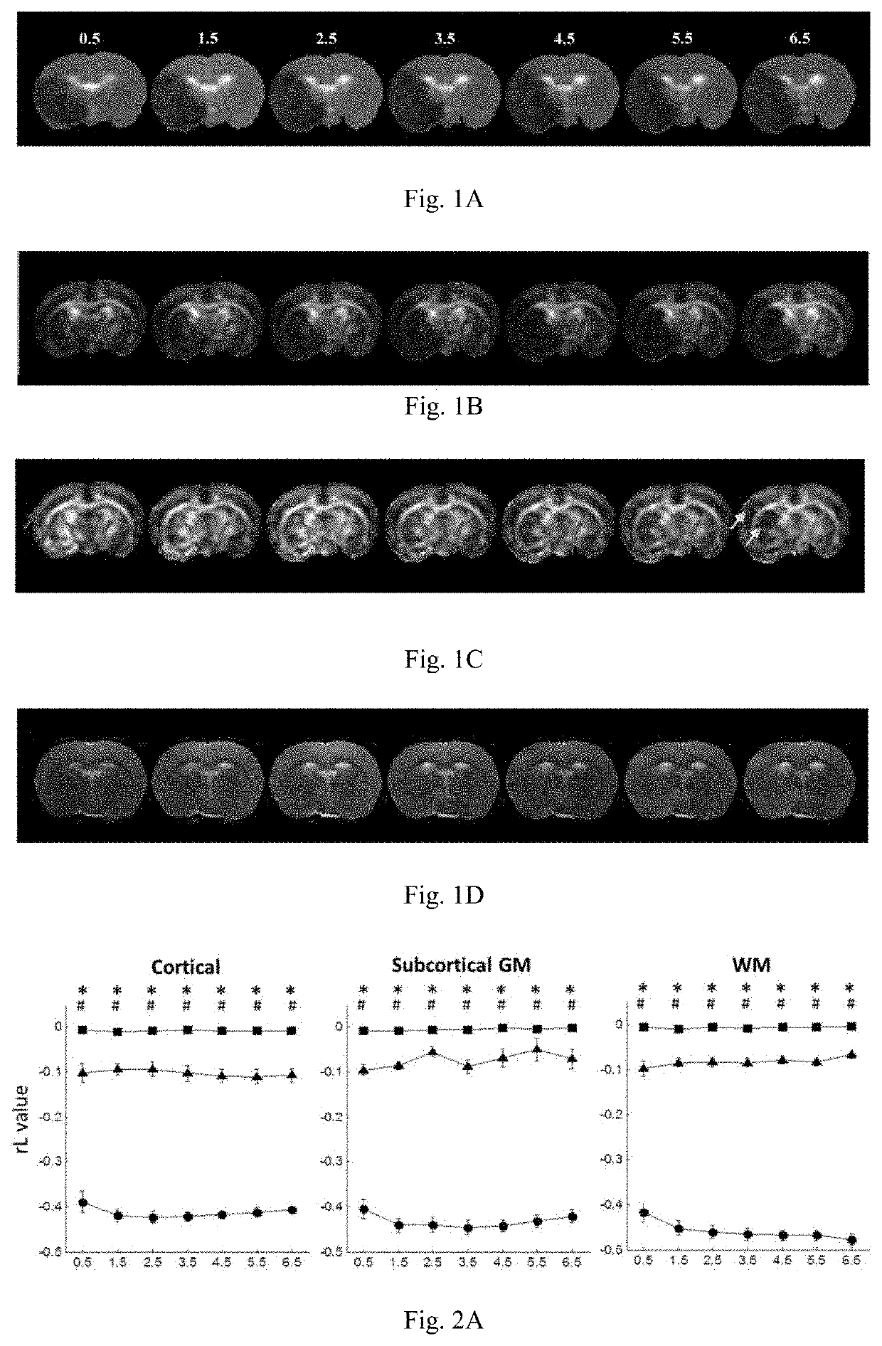
[0117]The experiment procedure was approved by the local Institute of Animal Care and Utilization Committee. Eleven male Sprague-Dawley rats (250-300 g; National Laboratory Animal Center, Taiwan) were prepared by permanent occlusions of unilateral MCA using the intra-luminal suture as proposed by Chiang et al. (Chiang T, Messing R O, Chou W-H. Mouse model of middle cerebral artery occlusion. Journal of visualized experiments: JoVE 2011). Three of eleven rats died within 6.5 hours after MCAo and were therefore excluded from the subsequent analysis. One of the eight included rats died after the 24-hour imaging.

Magnetic Resonance Imaging

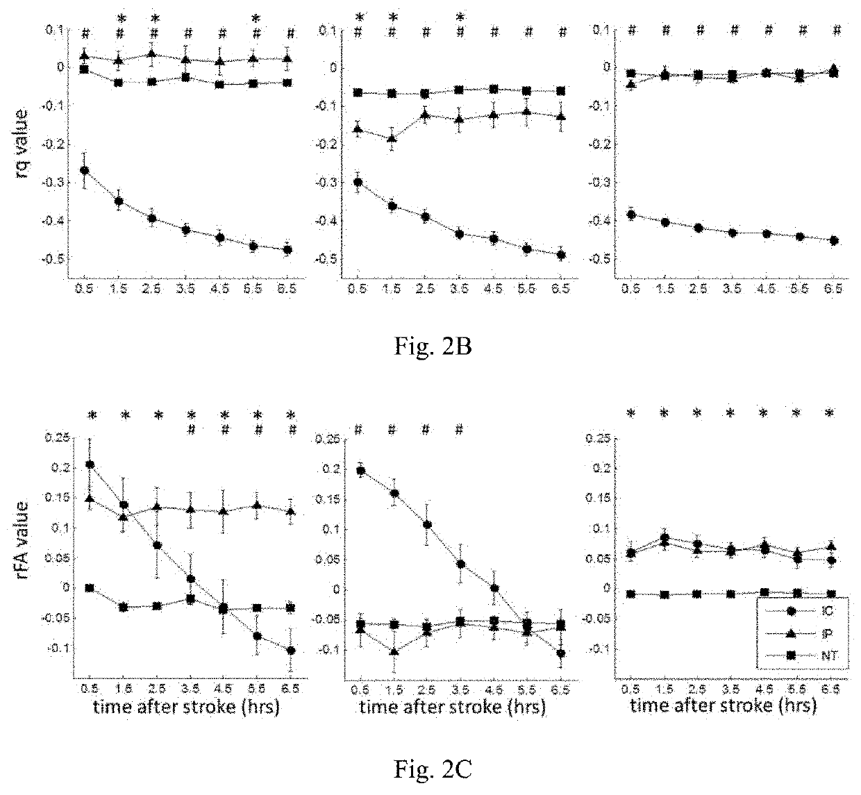
[0118]All MRI animal experiments were performed in a 7T scanner (PharmaScan 70 / 16; Bruker, Germany). The rats were maintained under anesthesia using 1.5-2% isoflurane with an oxygen flow of 1 L / min. Rectal temperature were kept at 37° C. by infusing warm air through the magnet bore. T2-weighted imaging (T2WI), dif...


PUM


Abstract

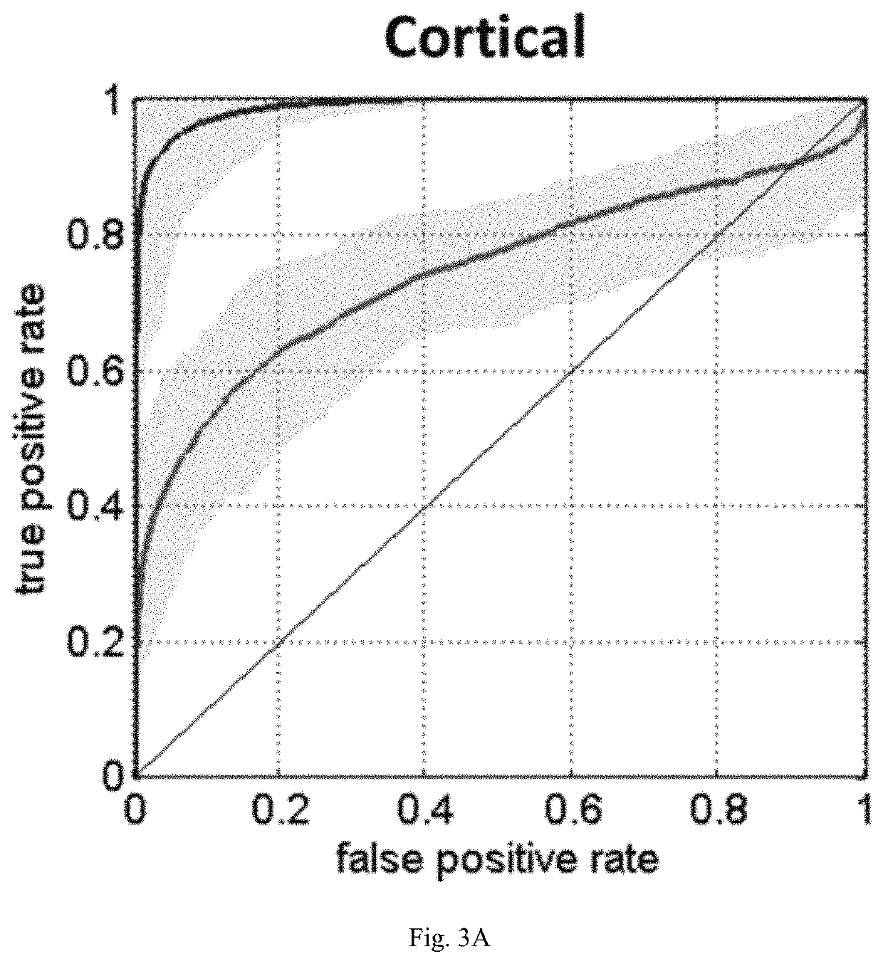
The invention relates to a method for determining ischemic status. The method comprises acquiring magnetic resonance diffusion tensor matrices and obtaining a relative decrease of diffusion magnitude due to the ischemic status from the magnetic resonance diffusion tensor matrices. The invention also relates to a method for assessing stroke onset time. The method comprises acquiring magnetic resonance diffusion tensor matrices and obtaining a relative decrease of pure anisotropy due to stroke from the magnetic resonance diffusion tensor matrices.

Description

BACKGROUND OF THE INVENTION1. Field of the Invention[0001]The invention relates to uses of magnetic resonance imaging (MRI). Particularly, the invention relates to a method for determining ischemic status or assessing stroke onset time of a brain region by magnetic resonance diffusion tensor matrices.2. Description of the Related Art[0002]Clinically, the golden hour of acute ischemic stroke is from the first minutes after onset to the next 3-4.5 hours. Hence, identification and differentiation of infarct core (the necrosis region due to ischemia) and reversible ischemic penumbra by non-invasive examination within a shortest time is critical for assessing the necessity of surgical intervention.[0003]On the other hand, in a significant proportion of patients, the exact time of stroke onset is unknown. Up to about one-quarter of the acute ischemic stroke patients, they either wake up with stroke symptoms or are found without conscious, and the stroke onset time cannot be ascertained. A...

Claims


Application Information

Patent Timeline
Patent Type & Authority Patents(United States)
IPC IPC(8): A61B5/00A61B5/055G01R33/48G06T7/00G01R33/563
CPCA61B5/055A61B5/0042A61B5/7246A61B5/7264G01R33/4806G06T7/0012G01R33/56341G06T2207/10088G06T2207/30016G16H50/20
Inventor CHEN, CHENG-YUCHUNG, HSIAO-WENKUO, DUEN-PANGLU, CHIA-FENGKAO, YU-CHIEH JILL
Owner TAIPEI MEDICAL UNIV
Learn More