Methods for protein transfer

a protein and protein technology, applied in the field of protein transfer, can solve the problems of difficult expressing more than a costimulator (or coinhibitor), cumbersome and time-consuming transfection process, and often poorly transfectable apcs, including tumor cells

Inactive Publication Date: 2002-03-28
TYKOCINSKI MARK L
View PDF0 Cites 0 Cited by
  • Summary
  • Abstract
  • Description
  • Claims
  • Application Information

AI Technical Summary

Problems solved by technology

For example, APCs, including tumor cells, are often poorly transfectable.
In addition, transfection proceedings are cumbersome and time-consuming.
Furthermore, expressing more than a costimulator (or coinhibitor) is difficult.
These and other issues have impeded the widespread application of gene therapy for APC and tumor cell engineering.
This method has significant limitations, including its dependence on covalent modifications that could perturb multiple proteins on cell surfaces.

Method used


Image

  • Methods for protein transfer
  • Methods for protein transfer
  • Methods for protein transfer

Examples


example 1

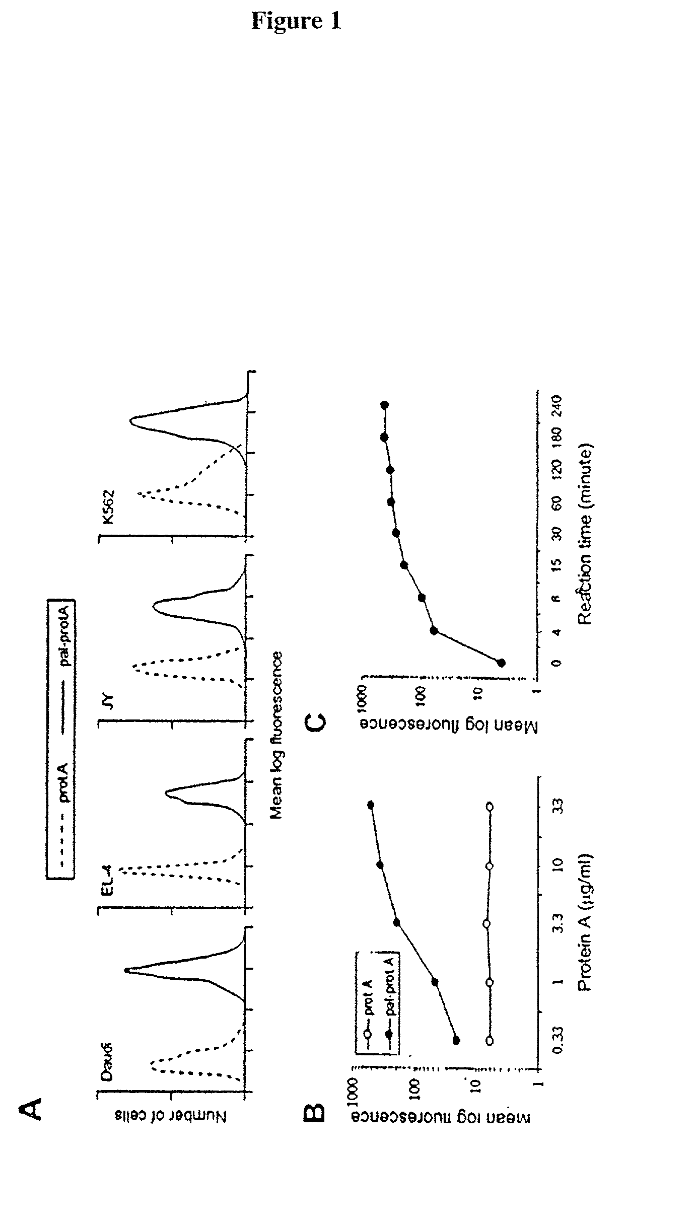
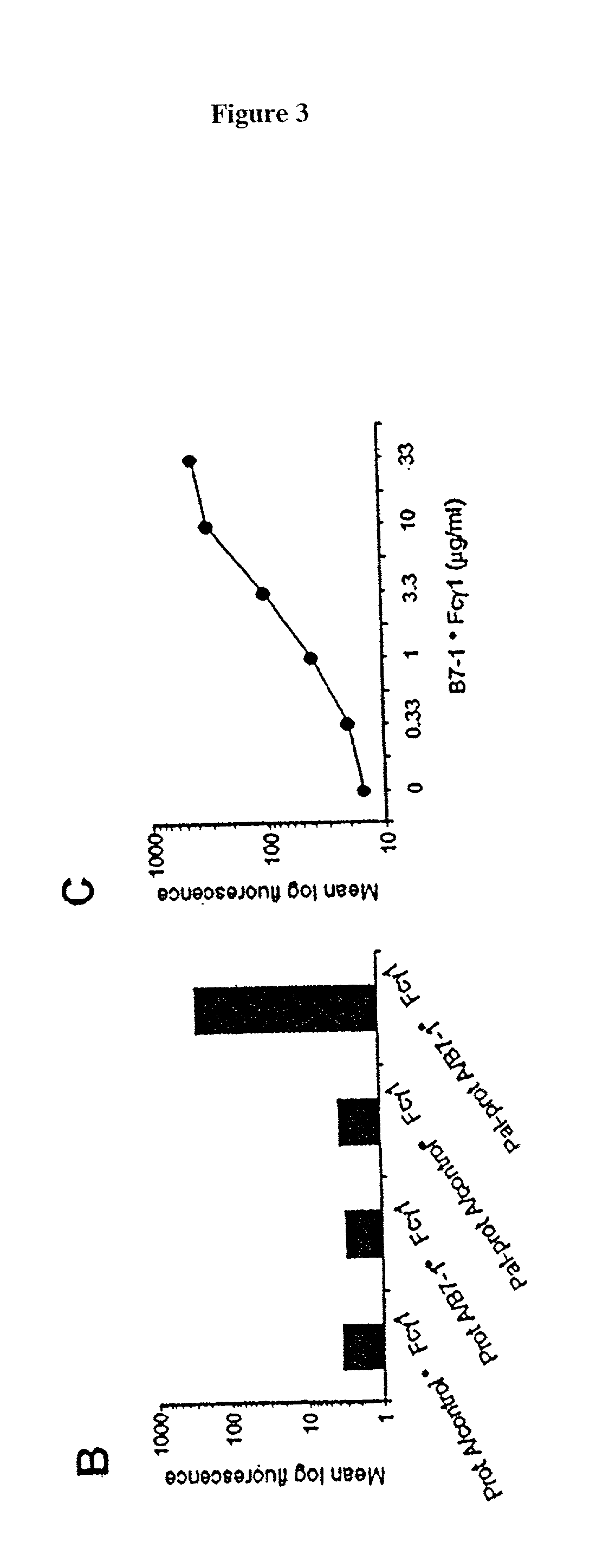
[0042] The following example demonstrates a method for transferring a B7-1-Fc.gamma..sub.1 fusion protein to a cell using palmitated protein A.

[0043] Palmitation of protein A

[0044] Recombinant protein A (Calbiochem, La Jolla, Calif.) was derivatized with the N-hydroxysuccinimide ester of palmitic acid (Sigma, St. Louis, Mo.) as described by Kim and Peacock, J. Immunol Methods, 158:57 (1993). Briefly, a stock solution of the N-hydroxysuccinimide ester of palmitic acid was made, as was a solution containing protein A in a concentration of about 1.5 mg / ml. The solutions were mixed in a ratio of about 10 .mu.g ester per ml protein and incubated at room temperature with constant mixing for about 18 h. The lipid-derivatized protein A was purified as described by Huang, et al., J. Biol. Chem., 225:8015 (1980) using a 30-ml Sephadex G-25 (Sigma) column. The protein product, referred to herein as "pal-prot A", was quantitated using a bicinchoninic acid kit (Bio-Rad, Richmond, Calif.), filter...

example 2

[0070] The effect of temperature on membrane-incorporated protein A was studied; the transferred protein must remain cell-bound in vivo in order to prime T-cells, which requires stable engagement of costimulators for at least several hours. It was determined that the reaction temperature at which a lipidated protein is transferred to the cell membrane has a major impact on long-term retention of the protein on the membrane. Protein transfer reactions were performed at 4.degree. C., 25.degree. C. or 37.degree. C.; palmitated protein A was transferred onto K562 cells. An hB7-1.multidot.Fc was .sup.125I-labeled and transferred to the protein A-coated cells in the manner described in Example 1. To prevent interference of endocytosis likely to occur at temperatures above 4.degree. C., the cells were treated with the metabolic inhibitors sodium azide and 2-deoxyglucose prior to the transfer reaction.

[0071] To determine the long-term retention of the transferred protein on the cell membran...

example 3

[0072] C3H / HeN mice, purchased from Harlen (USA), Indianapolis, were inmmunized with a cell vaccine generated from the T-50 cell line, obtained from Avranham Hochberg, Hadassah University Hospital. The vaccine was prepared following the procedure generally outlined in Example 1, using palmitated protein A and mB7-1.multidot.Fc, m4=1BBL.multidot.Fc, and hCD40L.multidot.Fc fusion proteins. Basically, the cells were coated with the lipidated protein A at 37.degree. C. at a ratio of 40 .mu.g protein A per 40.times.10.sup.6 cells. The cells were then incubated at 4.degree. with an equal mixture of the three fusion proteins at a ratio of 20 .mu.g total protein per 4.times.10.sup.7 cells. The cell vaccine was injected into the mice subcutaneously at a dose of 10.sup.6 cells per injection. The injections were given once a week and continued for three weeks. One week after the last injection, the animals were challenged with 10.sup.6 wild-type T-50 tumor cells, injected intradermally on the ...


PUM

PropertyMeasurementUnit
concentrationaaaaaaaaaa
concentrationsaaaaaaaaaa
densityaaaaaaaaaa

Abstract

Methods for transferring one or more proteins to a cell are disclosed. The protein or proteins to be transferred are in the form of a fusion protein, and contain at least one domain encoding for a protein or peptide having trans signaling and / or adhesion function. The fusion protein is transferred to a cell by binding to a lipidated protein, which has been incorporated into the cell membrane. Methods for using cells which have undergone protein transfer according to the present methods are also disclosed. This includes use in a cancer vaccine, use for treatment of cancer or autoimmune disease, and use in determining costimulator threshold levels.

Description

[0001] The present invention relates to novel methods for transferring one or more proteins to a cell. In addition to other applications, the methodology is useful in the treatment of cancer and autoimmune diseases, and for determining costimulator activation thresholds and cooperative interactions among costimulators.BACKGROUND INFORMATION[0002] T-cells, including cytotoxic T-lymphocytes (CTLs), are a critical component of effective human immune responses to tumors, viral infections and other infectious diseases. T-cells destroy neoplastic or virally infected cells through recognition of antigenic peptides presented by MHC class I molecules on the surfaces of target cells. Activation of T-cells is dependent upon coordinate signaling through antigen receptors and costimulator receptors on T-cell surfaces. Many mechanisms contribute to the escape of tumor cells and virally infected cells from immune surveillance. One of the mechanisms is that these cells lack the costimulatory molecu...

Claims


Application Information

Patent Timeline
Patent Type & Authority Applications(United States)
IPC IPC(8): C12N5/07A61K35/12A61K38/00A61K39/00A61P35/00A61P37/02C07K14/705C12N5/00C12N5/02C12N5/078C12N5/09
CPCA61K35/12A61K39/0011A61K2039/5158A61K2039/55516A61K2039/6031C07K14/70532C07K14/70575C07K2319/00C07K2319/30C12N5/0006C12N2501/51C12N2501/58A61P35/00A61P37/02A61K39/001129A61K39/001166A61K2039/80
Inventor TYKOCINSKI, MARK L.CHEN, AOSHUANGZHENG, GUOXING
Owner TYKOCINSKI MARK L
Why Eureka
Try Eureka