Methods for modeling infectious disease and chemosensitivity in cultured cells and tissues

a technology of infectious diseases and cultured cells, applied in the field of microbiology, infectious diseases and drug toxicity, can solve the problems of inability to model the best conditions faced by tissue culture models, limited conclusions about the interaction between pathogens and host epithelium using cultured epithelial cells, and many of the characteristics of animal models fail to mimi

Inactive Publication Date: 2004-09-09
THE GOVERNMENT OF THE UNITED STATES OF AMERICA AS REPRESENTED BY THE DEPT OF VETERANS AFFAIRS +1
View PDF2 Cites 4 Cited by
  • Summary
  • Abstract
  • Description
  • Claims
  • Application Information

AI Technical Summary

Problems solved by technology

Some types of tissue culture models of infectivity are not the best models of the conditions faced in vivo by pathogens.
Conclusions drawn about the interaction between pathogens and the host epithelium using cultured epithelial cells may be limited due to the dedifferentiation of these cells during conventional cell culture.
Likewise, many of the characteristics of animal models fail to mimic the human disease, and animal models present a complex system in which many variables cannot be controlled.
Accordingly, since certain in vitro nor in vivo assays do not replicate the complex environment encountered by pathogens during the natural course of human infection, the information gained from these studies may be limited.
Specifically, tissue culture models of infectivity are not exact models of the conditions faced in

Method used


Image

  • Methods for modeling infectious disease and chemosensitivity in cultured cells and tissues
  • Methods for modeling infectious disease and chemosensitivity in cultured cells and tissues
  • Methods for modeling infectious disease and chemosensitivity in cultured cells and tissues

Examples


Embodiment Construction

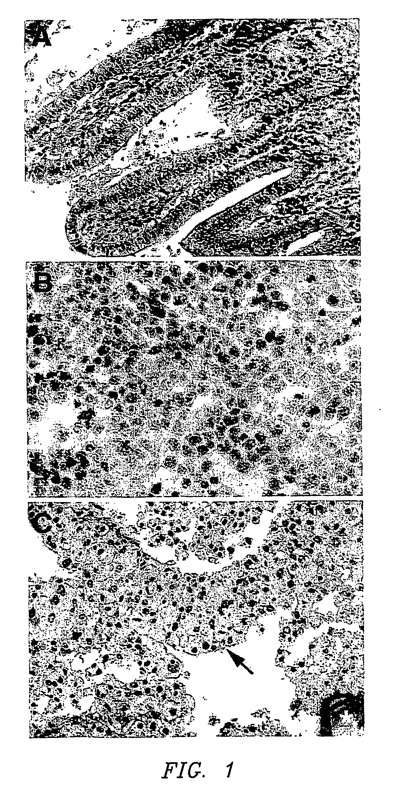
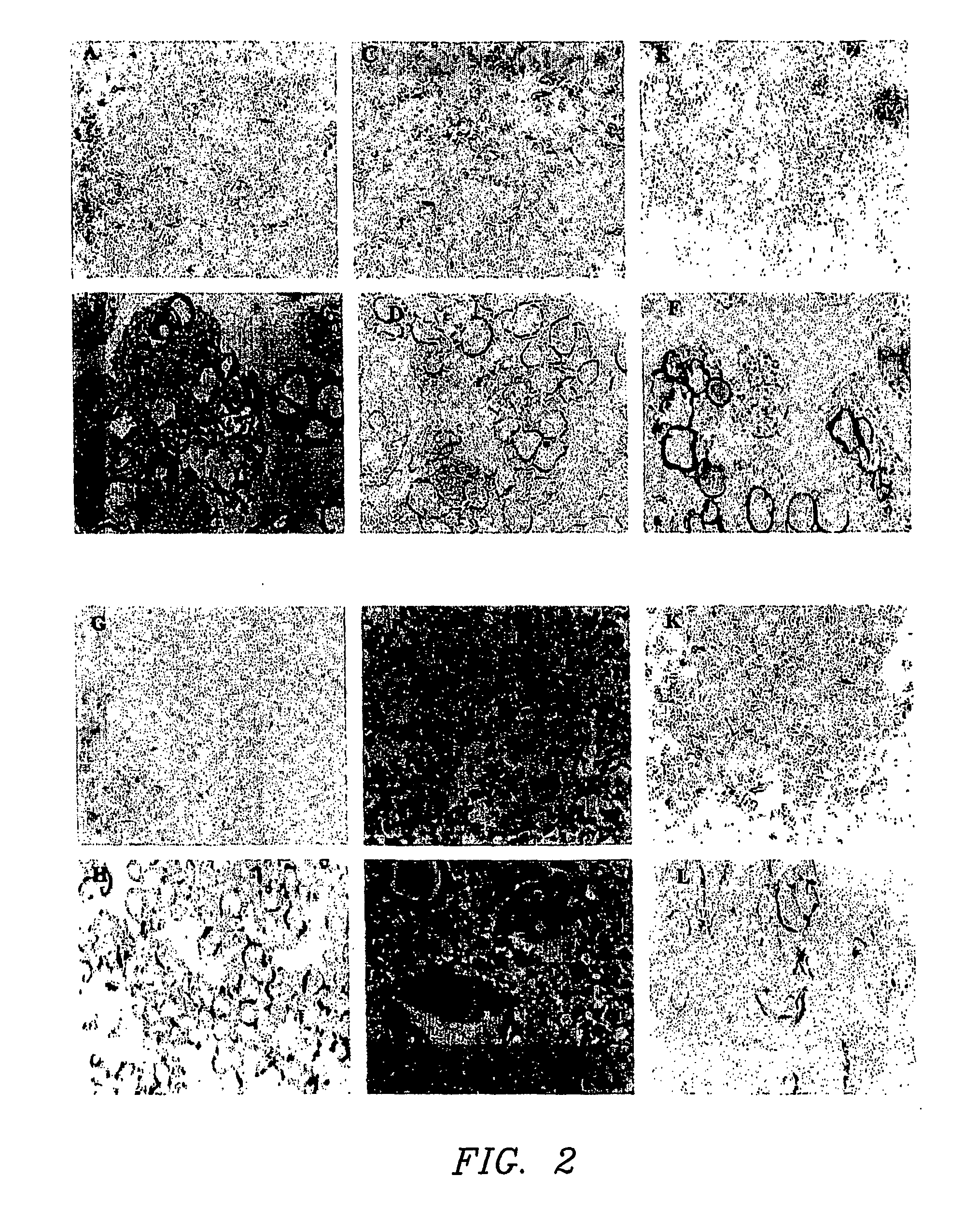
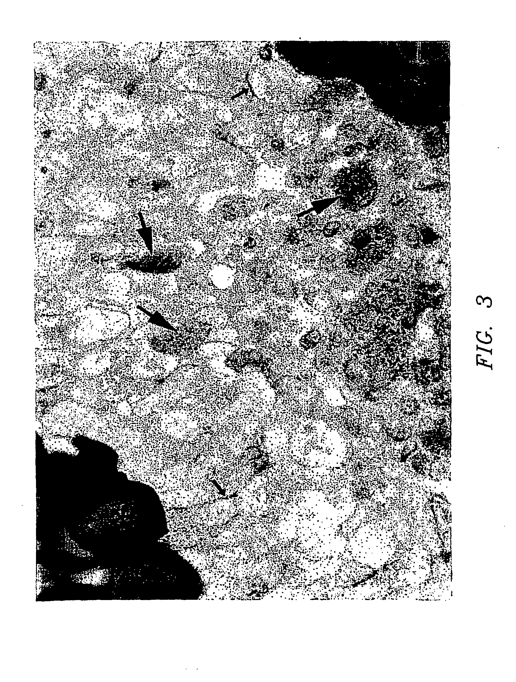
[0023] The present invention relates to the growth of cells in a suspension culture that due to low shear and low operational turbulence minimizes mechanical cell damage and allows cells to associate into three-dimensional structures, thereby promoting cellular differentiation. In an aspect of the invention, the three-dimensional cell structures (or 3-D aggregates) are used to study microbial infectivity from the perspective of the host-pathogen interaction. In another aspect of the invention, the three dimensional cell structures are used to study the chemosensitivity of the 3-D tissue mass to chemotherapeutic agents and toxins.

[0024] The present invention further relates to the use of a bioreactor for the growth of cells in a suspension culture, wherein the shear levels are defined. Specifically, cell culture is carried out in the bioreactor under conditions of applied shear, Coriolis forces applied, with cells falling at a terminal velocity. This tissue culture approach permits t...


PUM

PropertyMeasurementUnit
Fractionaaaaaaaaaa
Timeaaaaaaaaaa
Timeaaaaaaaaaa

Abstract

The present invention provides methods for utilizing a form of optimized suspension culture to examine the infectivity of pathogenic organisms and agents in human cells and tissues. Also provided are methods using a rotating wall vessel to predict chemosensitivity of cells and tissues to toxins and chemotherapeutic agents. These culture conditions potentiate spatial colocalization and three-dimensional assembly of individual cells into large aggregates which more closely resemble the in vivo tissue equivalent. In this environment, dissociated cells can assemble and differentiate into macroscopic tissue aggregates several millimeters in size. These culture conditions allow for better cellular differentiation and formation of three-dimensional cellular aggregates, more efficient cell-to-cell interactions, the in in vivo-like exchange of growth factors and greater molecular scaffolding facilitating mechanical stability for cells. The suspension culture system offers a new approach for studying microbial infectivity from the perspective of the host-pathogen interaction and also for analyzing chemosensitivity to toxins and chemotherapeutic agents.

Description

[0001] This application claims priority to U.S. Provisional Application Serial No. 60 / 282,007, which is fully incorporated by reference herein.[0003] 1. Technical Field of the Invention[0004] The present invention relates generally to the fields of microbiology, infectious diseases and drug toxicity. More specifically, the present invention relates to methods of modeling infectious disease and chemosensitivity with three-dimensional suspension of mammalian cells using a bioreactor.[0005] 2. Description of the Prior Art[0006] An essential feature of the pathogenicity of microorganisms is their ability to infect or colonize host epithelial cells and cells in other tissues. The terms "infect" and "colonize" are used interchangeably herein, and encompass adherence to cells, invasion, survival within, and damage or destruction of the cells. Initial sites of bacterial infection include the epithelia of the intestine, kidney, lung and bladder. There is a need to investigate how bacterial a...

Claims


Application Information

Patent Timeline
IPC IPC(8): C12Q1/10C12Q1/68G01N33/50
CPCC12Q1/10G01N33/5005G01N2800/52G01N33/5088G01N33/5014
Inventor HAMMOND, TIMOTHY GRANTNICKERSON, CHERYL ANNE
Owner THE GOVERNMENT OF THE UNITED STATES OF AMERICA AS REPRESENTED BY THE DEPT OF VETERANS AFFAIRS
Why Eureka
Try Eureka