Nuclear imaging system using scintillation bar detectors and method for event position calculation using the same

a technology of scintillation bar and detector, applied in the field of nuclear medicine, can solve the problem that the performance of bar detectors as designed in the prior art is insufficient for medical imaging applications, and achieve the effect of improving spatial resolution

Inactive Publication Date: 2005-01-13
SIEMENS MEDICAL SOLUTIONS USA INC
View PDF5 Cites 112 Cited by
  • Summary
  • Abstract
  • Description
  • Claims
  • Application Information

AI Technical Summary

Benefits of technology

According to another aspect of the invention, a bar detector gamma camera is provided, which utilizes an improved positioning algorithm

Problems solved by technology

However, the performance of bar detectors as designed in the pr

Method used


Image

  • Nuclear imaging system using scintillation bar detectors and method for event position calculation using the same
  • Nuclear imaging system using scintillation bar detectors and method for event position calculation using the same
  • Nuclear imaging system using scintillation bar detectors and method for event position calculation using the same

Examples


Embodiment Construction

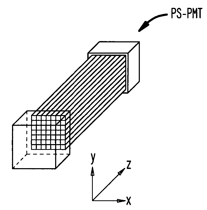
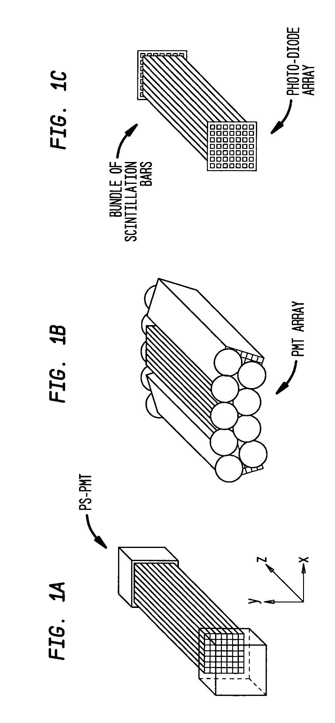
Referring to FIGS. 1(a)-1(c), according to one preferred embodiment of the invention, a bar detector module is constructed by bundling together a number of scintillation crystal bars in a two-dimensional array in the X-Y direction as shown. A nuclear medical imaging gamma camera then can be constructed by aggregating a number of modules together in a preferred geometry to establish a desired field of view for medical imaging applications. In the examples illustrated, the bars are arranged in a 7×7 square; however, many other configurations are possible and contemplated according to the invention. The bar detector module is optically coupled at each end to at least one photosensor. As shown in FIG. 1(a), each module may be optically coupled to a position-sensitive photomultiplier tube (PS-PMT). The PS-PMT is a known photosensor that has the ability to identify the X-Y location of light photons incident on its detection surface and produce an electric signal indicative of such X-Y lo...


PUM


Abstract

A gamma camera having a scintillation detector formed of multiple bar detector modules. The bar detector modules in turn are formed of multiple scintillation crystal bars, each being designed to have physical characteristics, such as light yield, to achieve a sufficient spatial resolution for nuclear medical imaging applications. According to another aspect of the invention, the bar detector modules are arranged in a three-dimensional array, where each module is made up of a two-dimensional array of bar detectors with at least one photosensor optically coupled to each end of the module. Such a camera can be used for both PET (coincidence) and single photon imaging applications. According to another aspect of the invention, a bar detector gamma camera is provided, which utilizes an improved positioning algorithm that greatly enhances spatial resolution in the z-axis direction (i.e., the direction along the length of the scintillation crystal bar).

Description

BACKGROUND OF THE INVENTION 1. Field of the Invention The present invention generally relates to nuclear medicine, and systems for obtaining nuclear medicine images of a patient's body organs of interest. In particular, the present invention relates to a novel detector configuration for nuclear medical imaging systems that are capable of performing either positron emission tomography (PET) or planar and single photon emission computed tomography (SPECT). 2. Description of the Background Art Nuclear medicine is a unique medical specialty wherein radiation is used to acquire images that show the function and anatomy of organs, bones or tissues of the body. Radiopharmaceuticals are introduced into the body, either by injection or ingestion, and are attracted to specific organs, bones or tissues of interest. Such radiopharmaceuticals produce gamma photon emissions that emanate from the body. One or more detectors are used to detect the emitted gamma photons, and the information coll...

Claims


Application Information

Patent Timeline
IPC IPC(8): G01T1/00G01T1/20G01T1/202
CPCG01T1/202G01T1/2018G01T1/20182G01T1/20187
Inventor JOUNG, JINHUNCHOWDHURY, SAMIRENGDAHL, JOHN C.
Owner SIEMENS MEDICAL SOLUTIONS USA INC
Why Eureka
Try Eureka