System and method of anatomical modeling

an anatomical modeling and system technology, applied in the field of anatomical and pathological modeling, can solve the problems of preventing real-time implementation, computationally demanding, and boolean operations for medical objects, but not particularly meaningful, and processing the three-dimensional data directly from the original medial image,

Inactive Publication Date: 2005-01-27
AGENCY FOR SCI TECH & RES
View PDF17 Cites 219 Cited by
  • Summary
  • Abstract
  • Description
  • Claims
  • Application Information

AI Technical Summary

Benefits of technology

In essence, this aspect of the invention allows information regarding the representation of the anatomical structure to be conveniently stored and compiled. It provides a solution for the problem of surface reconstruction from parallel-and non-parallel cross-sections with visually smooth surfaces rendered, wherein a novel joint construction approach deals with the difficult branching problem. The reconstructed geometrical surface allows visualization and manipulation flexibly performed, and the computational demand is within a standard PC.
In particular, the second and third aspects of the invention provide for pathology modeling in respect of vasculature. This aspect of the invention further allows models of pathology to be developed, which incorporate patient specific data, and hence are more suitable for and characteristic of patient specific applications.

Problems solved by technology

This can provide an accurate representation of the anatomical structure, but the model is usually intended for specific cases and cannot be applied generally.
Further, processing the three-dimensional data directly from original medial images, such as medical resonance images (MRI), computerized tomography (CT) or ultrasound scan images, is computationally demanding.
This size will prohibit the implementation in real-time.
Boolean operations for medical objects, however, are not particularly meaningful.
A problem with boundary representations is that they are usually computationally expensive.
However, they do have deficiencies, particularly in medical applications, where model reconstruction from various modalities of images is an important issue.
Another problem that needs to be addressed in geometrical modeling is that of surface reconstruction from cross-sections.
It has been found that a surface reconstructed from parallel cross-contours may not be good for visualization, especially if the central axis of the human vasculature is slanted with the slice.
Further, if the scanned medical image includes different kinds of human anatomy, to date a suitable automated process for reconstructing the desired component of the image has not been developed.
The volume-based approach is difficult to use in cases where the cross-sectional shape varies between planes and where the cross sections are non-parallel.
Further, it is difficult to constrain the aspect ratios of the generated triangles, since the vertices are defined by the positions of the cross sections.
This can lead to poor quality surface displays if Gouraud shading is used.
In this regard, much effort is also required to detect and correct special cases where the triangulation of complex shapes might otherwise fail.
One problem with existing surface reconstruction methods, however is that they do not adequately allow branching junctions to be reconstructed.
In this regard, the modeling of the branching junction is a challenging issue.

Method used


Image

  • System and method of anatomical modeling
  • System and method of anatomical modeling
  • System and method of anatomical modeling

Examples


Embodiment Construction

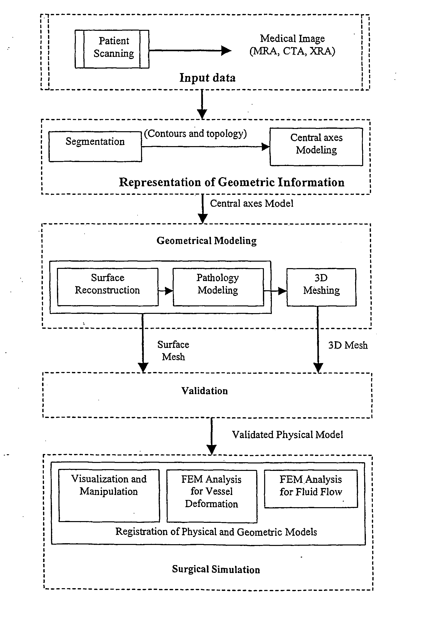
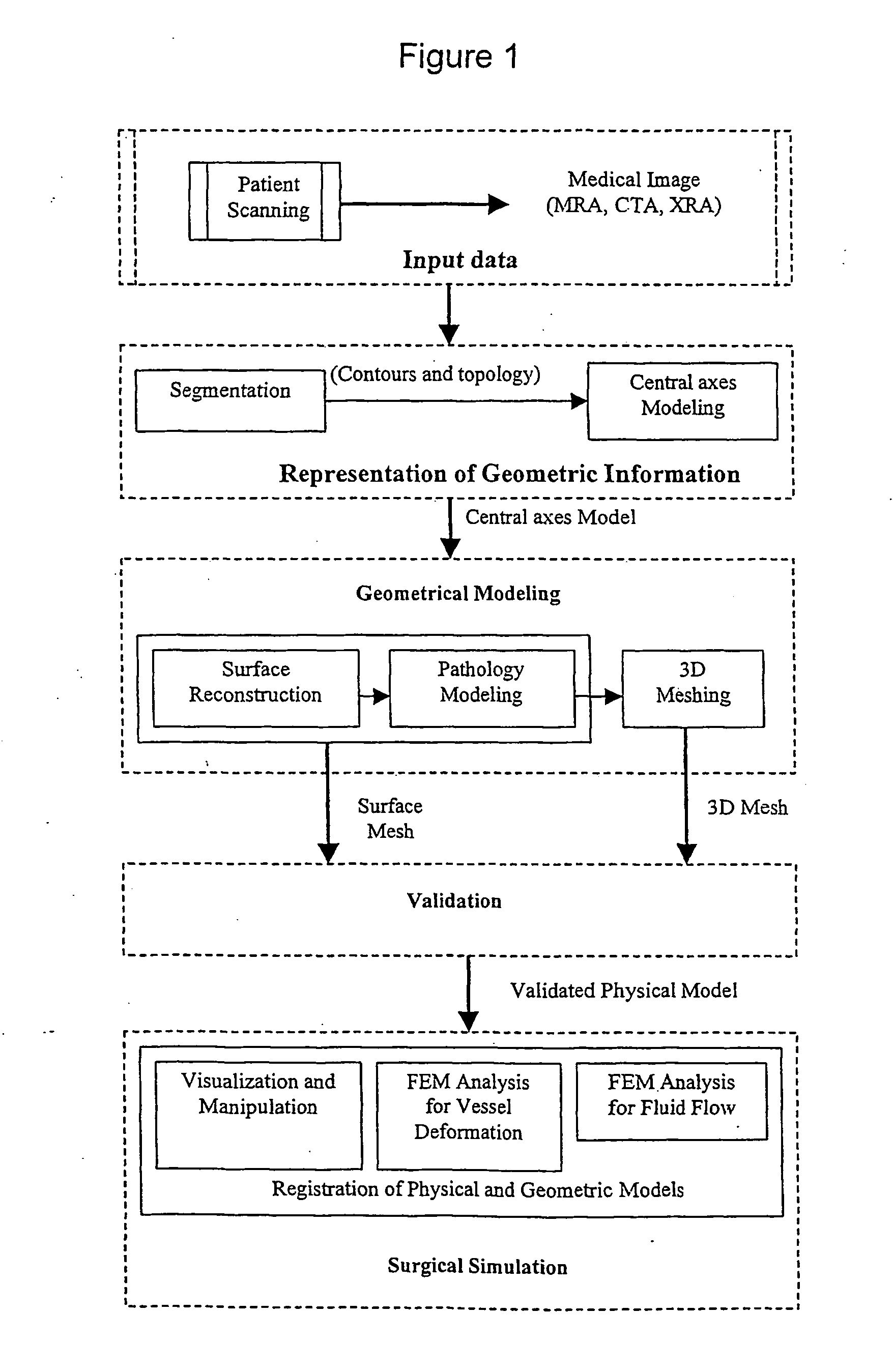
With reference to FIG. 1, a flow chart is provided which indicates the general steps taken in regard to creating a human anatomy model for surgical simulation.

The first stage is that of obtaining input data for creating the model. This may be achieved by scanning the relevant patient using, for example, MRI, CT, Ultrasound, X-ray rotational angiography (XRA) and obtaining the appropriate medical volume images therefrom.

It is also to be appreciated that where the present invention is being utilised in an operative situation, such as image-guided surgery, the input data may be a combination of pre-operatively acquired images and intra-operatively acquired images.

Once this input data has been obtained, the next stage is to derive a representation of the geometric information. Hence, the topological and geometrical information like contours, radii and central axes are extracted from the medical images. With this information segmentation is performed in order to obtain an appropri...


PUM


Abstract

Methods of modeling anatomical structures, along with pathology including the vasculature, spine and internal organs, for visualization and manipulation in simulation systems. A representation of on the human vascular network is built up from medical images and a geometrical model produced therefrom by extracting topological and geometrical information. The model is constructed using topological and geometrical information. The model is constructed using segments containing topology structure information, flow domain information contour domain information and skeletal domain information. A realistic surface is then applied to the geometric model, by generating a trajectory along a central axis of the geometric model, conducting moving trihedron modeling along the generated trajectory and then creating a sweeping surface along the trajectory. A novel joint reconstruction approach is also proposed whereby a part surface sweeping operation is performed across branches of the joint and then a surface created over the resultatn holes therebetween. A 3-D mesh may also be generated, based upon this model, for finite element analysis and pathology creation.

Description

FIELD The present invention relates to anatomical and pathological modeling as utilised in virtual reality simulation environments, scientific visualization, computer aided geometric design and finite element analysis. More particularly the present invention relates to human anatomical and pathological modeling for visualization and manipulation in surgical simulation systems such as for the purposes of surgery planning, training and education. BACKGROUND Medical simulators have significant potential in reducing the cost of health care through improved training and better pretreatment planning. Further, image guided procedures, such as vascular catheterization, angioplasty and stent placement are especially suited for simulation because it is possible to place the physician at a distance from the operative site, thereby remotely manipulating surgical instruments and viewing the procedure on video monitors. For real-time and useful simulation of interventional procedures, such as ...

Claims


Application Information

Patent Timeline
Patent Type & Authority Applications(United States)
IPC IPC(8): G06T17/00
CPCG06T17/00
Inventor CHEN, XUESONGCHUI, CHEE KONGNOWINSKI, WIESLAW T.
Owner AGENCY FOR SCI TECH & RES
Why Eureka
Try Eureka