
Optical in vivo analyte probe using embedded intradermal particles

an in vivo analyte and probe technology, applied in the field of in vivo detection and/or quantification of analytes, can solve the problems of historic difficulty of sers detection of glucose in vivo using prior art methods, less appropriate uv resonance enhanced raman for in vivo detection applications,

Inactive Publication Date: 2005-02-03
MARBLIA LTD +1
View PDF10 Cites 67 Cited by
  • Summary
  • Abstract
  • Description
  • Claims
  • Application Information

AI Technical Summary

Benefits of technology

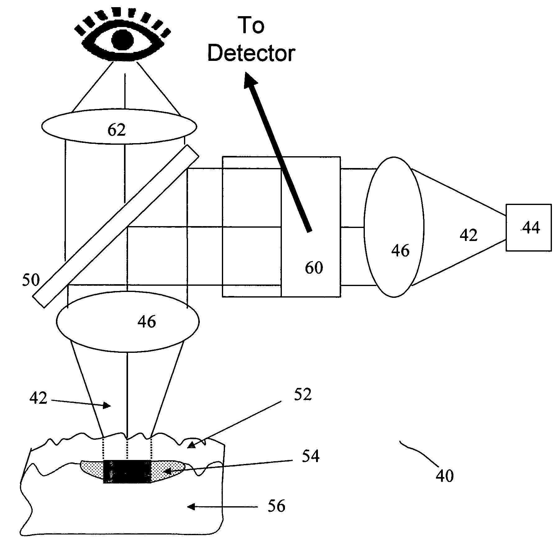
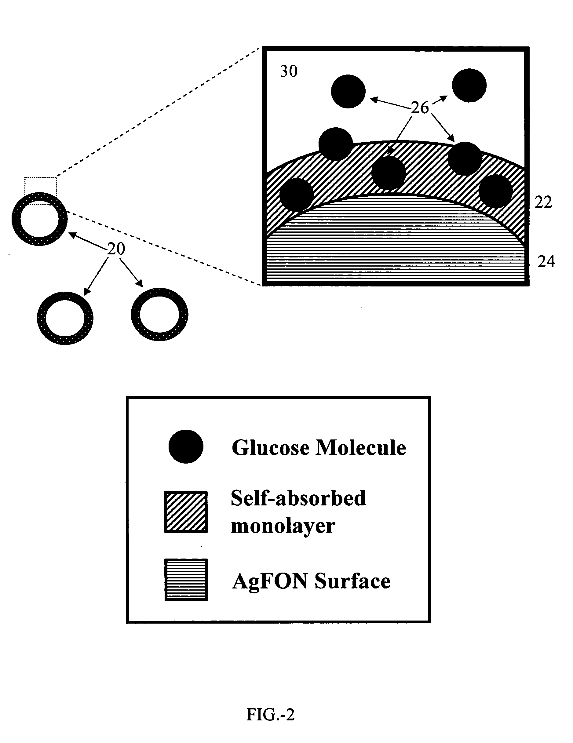
The present invention provides for in vivo optical detection of analytes (e.g., glucose) by embedding small particles within the dermis of the skin. The small particles are made sensitive to the analyte of interest by construction. For example, silver-coated nanoparticles further covered with a self-assembled molecular monolayer are suitably sensitized for the detection of glucose. The particles embedded within the dermis are preferably probed by surface enhanced Raman spectroscopy, although other optical probe techniques can be employed.

Problems solved by technology

UV resonance enhanced Raman is less appropriate for in vivo detection applications because of limitations in the tissue transparency window.
The historic difficulty of SERS detection of glucose in vivo using prior art methods may be attributable to the weak or nonexistent binding of glucose to bare silver surfaces.

Method used


Image

  • Optical in vivo analyte probe using embedded intradermal particles
  • Optical in vivo analyte probe using embedded intradermal particles
  • Optical in vivo analyte probe using embedded intradermal particles

Examples


Embodiment Construction

1. Selective Enhanced Glucose Detection

Detection of glucose (or other analytes) using SERS presents two potential problems. Silver nano-particles (or other sensitized small particles) must be implanted in the human body to effectively make contact with glucose, and the nano-particles must be sensitized to glucose, which generally does not readily adhere to silver surfaces. The present invention solves the first problem by applying well-known tattooing techniques for implantation of sensitized particles.

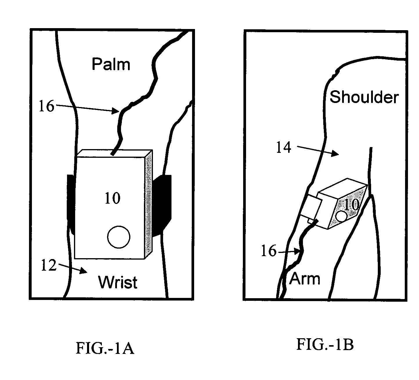
Referring to FIG.1A and FIG.1B, a monitor 10 according to the present invention may advantageously be worn on the inner wrist 12 as shown in FIG.1A, on the inner upper arm 14 as shown in FIG.1B, or, alternatively, on some other portion of the body where close and consistent skin contact may be maintained. For the examples shown in FIG.1A and FIG.1B, contact is made to skin on the inner part of the wrist or arm, respectively. A fiber cable 16 connects the monitor unit to a laser s...


PUM


Abstract

A system and method of in vivo detection and quantification of one or more analytes. Small particles comprising a surface-active monolayer coating are embedded in the dermis. The surface-active monolayer acts to pre-concentrate the analyte by adsorbing the analyte from bulk solution. The concentrated analyte is more readily detected and quantified by one or more spectroscopic methods such as Raman spectroscopy

Description

FIELD OF THE INVENTION The present invention relates generally to the field of in vivo detection and / or quantification of analytes. More specifically, the present invention relates to systems and methods for detecting, analyzing, and / or quantifying concentrations of compounds by measuring the spectral responses of sensitized particles embedded within tissue such as the skin. BACKGROUND OF THE INVENTION Raman spectroscopy is a powerful tool for revealing specific molecular signatures from a complex system. For glucose in vivo detection, a Raman excitation laser emitting in the near IR region of the spectrum can penetrate into tissues to probe the molecular vibration which can not be done by IR spectroscopy due to strong water absorption. However, Raman scattering is a nonlinear process and tends to have a very small cross section (i.e., approximately 10−30 cm−2 sr−1 molecule−1). Two known approaches for enhancing Raman scattering processes are surface enhanced Raman spectroscopy (S...

Claims


Application Information

Patent Timeline
Patent Type & Authority Applications(United States)
IPC IPC(8): A61B5/00
CPCA61B5/14532B82Y30/00A61B5/411
Inventor XIE, JINCHUN
Owner MARBLIA LTD
Learn More